The eye is a specialized sensory organ of photoreception. The eye is an easily accessible organ for local or systemic drug delivery.
Clinically, the eye can be considered to be composed of two segments:
1. Anterior segment – all structures from (and including) the lens forward.
2. Posterior segment – all structures posterior to the lens.
- The anatomical and physiological characteristics of the eye are described are outlined in this section.
- Structure of the eye; The eye can be divided into two compartments: the anterior and posterior segments.
- An internal cross section of an eye is shown in Fig.1
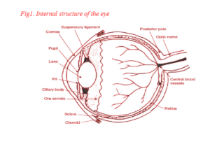
- Anterior segment; Externally, the anterior segment of eye is made up of cornea, conjunctiva, and sclera.
- Internally, it consists of anterior chamber, iris/pupil, posterior chamber, and ciliary body.
- The cornea, an optically transparent tissue that aids in refraction of light to the eye for focusing, is 1 mm thick at the periphery and 0.5 to 0.6 mm thick in the centre.
- It is composed of squamous and basal columnar epithelium, Bowman’s membrane, substantia propria (stroma), limiting lamina, and the endothelium.
- The conjunctiva is a thin, transparent, vascularised mucous membrane with an area of 18 cm2 covering the eye globe and the inner eyelids.
- It maintains the precorneal tear film and protects the eye. It produces mucus and lubricates the surface of the eye.
- It is made up of stratified columnar epithelium and lamina propria. The conjunctiva epithelium is divided into bulbar (covering the eyeball), fornix (covering the cornea), and palpebral (covering the eyelid) conjunctivae.
- The sclera, the white outer coat of the eyeball, provides structural integrity, size, and shape to the eye.
- There are three layers in the sclera, the anterior episclera, the middle scleral stroma, and the posterior lamina fusca.
- The sclera is composed of gel like mucopolysaccharides, elastic fibers, bundles of dense collagen fibrils, and fibroblasts.
- The iris is a diaphragm around the pupil (lens) and controls the amount of light entering the inner eye.
- The ciliary body is made up of ciliary muscles, which aid in accommodation.
- The anterior surface of the eye is constantly rinsed by tear fluid secreted at a flow rate of about 1 μL/min by the main lachrymal gland of the lachrymal apparatus.
- Tears eventually drain into the nasal cavity through the nasolachrymal ducts.
- Tear fluid contains mucin, lysozyme, lactoferrin, prealbumin, and serum proteins.
- It functions as an antibacterial lubricant and aids in draining out foreign substances.
- The normal volume of tear fluid is 5 to 10 μL.65
Posterior segment:
- Externally, the posterior segment consists of the optic nerve and associated vasculature, and internally, it consists of the lens, vitreous, and rear ocular tissues. Vitreous is a colorless medium
- Internal structure of the eye consisting of about 99 percent water, dissolved type II collagen, sodium hyaluronate, and proteoglycans.
- The retina is the inner nervous layer of the eye responsible for the sensory function of sight.
- The choroid is a dark brown vascular layer attached to the sclera and is believed to provide nourishment to the retina.
Basic Structure of the Eye
The eye has three layers or coats, three compartments and contains three fluids
1. The three coats of the eye are as follows:
(a) Outer fibrous layer:
• cornea
• sclera
• lamina cribrosa.
(b) Middle vascular layer (“uveal tract”):
• iris
• ciliary body – consisting of the pars
plicata and pars plana
• choroids.
(c) Inner nervous layer:
• pigment epithelium of the retina
• retinal photoreceptors
• retinal neurons.
2. The three compartments of the eye are as follows:
(a) Anterior chamber – the space between the cornea and the iris diaphragm.
(b) Posterior chamber – the triangular space between the iris anteriorly, the lens and zonule posteriorly, and the ciliary body.
(c) Vitreous chamber – the space behind the lens and zonule.
3. The three intraocular fluids are as follows:
(a) Aqueous humour – a watery, optically clear solution of water and electrolytes similar to tissue fluids except that aqueous humour has a low protein content normally.
(b) Vitreous humour – a transparent gel consisting of a three-dimensional network of collagen fibres with the interspaces filled with polymerised hyaluronic acid molecules and water. It fills the space between the posterior surface of the lens, ciliary body and retina.
(c) Blood – in addition to its usual functions, blood contributes to the maintenance of intraocular pressure. Most of the blood within the eye is in the choroid. The choroidal blood flow represents the largest blood flow per unit tissue in the body. The degree of desaturation of efferent choroidal blood is relatively small and indicates that the choroidal vasculature has functions beyond retinal nutrition. It might be that the choroid serves as a heat exchanger for the retina, which absorbs energy as light strikes the retinal pigment epithelium.