M Pharmacy Parmaceutics Project : APPROACHES TO COLON-SPECIFIC DRUG DELIVERY [Ceutics]

M Pharmacy Parmaceutics Project APPROACHES TO COLON-SPECIFIC DRUG DELIVERY [Ceutics]

M Pharmacy Project : APPROACHES TO COLON-SPECIFIC DRUG DELIVERY

APPROACHES TO COLON-SPECIFIC DRUG DELIVERY

In recent years, a large number of solid formulations targeting the lower parts of the GI tract, especially the colon, have been reported. These formulations may be broadly divided into four types, which are

  1. pH-dependent system designed to release a drug in response to change in pH,
  2. Time controlled ( or Time-dependent) system designed to release a drug after a predetermined time,
  3. Microbially-controlled system making use of the abundant enterobacteria in the colon,
  4. Enzyme-based systemsProdrug, and
  5. Pressure-dependent system making use of luminal pressure of the colon.

Among these, first three are most widespread formulation technologies being developed for pharmaceutical market.


pH-DEPENDENT SYSTEMS

Solid formulations for colonic delivery that are based on pH-dependent drug release mechanism are similar to conventional enteric-coated formulations but they differ in target site for delivery and therefore type of enteric polymers. In contrast to conventional enteric-coated formulations, colonic formulations are designed to deliver drugs to the distal (terminal) ileum and colon, and utilize enteric polymers that have relatively higher threshold pH for dissolution (Dew et al., 1982; Tuleu et al., 2001). Most commonly used polymers (Table 2) are derivatives of acrylic acid and cellulose. These polymers have ability to withstand an environment ranging from low pH (~1.2) to neutral pH (~7.5) for several hours. Apparently, it is highly desirable for pH-dependent colonic formulations to maintain their physical and chemical integrity during passage through the stomach and small intestine and reach the large intestine where the coat should disintegrate to release the drug locally. It should be however noted that GI fluids might pass through the coat while the dosage form transits through the small intestine. This could lead to premature drug release in the upper parts of GI tract and as a result loss of therapeutic efficacy may occur. One approach to overcome this problem is to apply

higher coating levels of enteric polymers; however, this also allows influx of GI fluids through the coat, and the thicker coats often rupture under the influence of contractile activity in the stomach. In general, the amount of coating required depends upon the solubility characteristics (solubility, dose/solubility ratio) of the drug, desired release profile and surface area of the formulation, and composition of the coating solution/dispersion.

Widely used polymers are methacrylic resins (Eudragits), which are available in water soluble and water-insoluble forms. Eudragit L and S are copolymers of methacrylic acid and methyl methacrylate. To overcome the problem of premature drug release, a copolymer of methacrylic acid, methyl methacrylate and ethyl acrylate (Eudragit® FS), which dissolves at a slower rate and at a higher threshold pH (7–7.5), has been developed recently. A series of in vitro dissolution studies with this polymer have highlighted clear benefits over the Eudragit® S polymer for colonic targeting (Rudolph et al., 2001).

M Pharmacy Parmaceutics Project APPROACHES TO COLON-SPECIFIC DRUG DELIVERY [Ceutics] M Pharmacy Project APPROACHES TO COLON-SPECIFIC DRUG DELIVERY pdf M Pharmacy Project APPROACHES TO COLON-SPECIFIC DRUG DELIVERY

Khan et al., (1999) prepared lactose-based placebo tablets and coated using various combinations of two methacrylic acid polymers, Eudragit® L100-55 and Eudragit® S100 by spraying from aqueous systems. The Eudragit® L100-55 and Eudragit® S100 combinations studied were 1:0, 4:1, 3:2, 1:1, 2:3, 1:4, 1:5 and 0:1. The coated tablets were tested in vitro for their suitability for pH dependent colon targeted oral drug delivery. The same coating formulations were then applied on tablets containing mesalazine as a model drug and evaluated for in vitro dissolution rates under various conditions. The disintegration data obtained for the placebo tablets demonstrate that disintegration rate of the studied tablets is depends on the polymer combinations used to coat the tablets, pH of the disintegration media and the coating level of the tablets. Dissolution studies performed on the mesalazine tablets further confirmed that the release profiles of the drug could be manipulated by changing the Eudragit® L100-55 and Eudragit® S100 ratios within the pH range of 5.5 to 7.0 in which the individual polymers are soluble respectively, and a coating formulation consisting of a combination of the two copolymers can overcome the issue of high GI pH variability among individuals. The results also demonstrated that a combination of Eudragit® L100-55 and Eudragit® S100

could be successfully used from aqueous system to coat tablets for colon targeted drug delivery and the formulation can be adjusted to deliver drug at any other desirable site of the intestinal region of the GIT on the basis of pH variability.

Colon targeted drug delivery systems based on methacrylic resins has described for insulin (Touitou and Rubinstein., 1986), prednisolone (Thomos., 1985), quinolones (Van Saene et al., 1986), salsalazine (Riley et al., 1987), cyclosporine (Kim et al., 2001), beclomethasone dipropionate (Levine et al., 1987) and naproxane (Hardy et al., 1987). pH-sensitive delivery systems are commercially available for mesalazine (5-aminosalicylic acid) (Asacol® and Salofalk®) and budesonide (Budenofalk® and Entocort®) for the treatment of ulcerative colitis and Crohn’s disease, respectively.


Table 2. Threshold pH of commonly used polymers    

Polymer 


Threshold pH
Eudragit® L100

Eudragit® S100

Eudragit® L 30D

Eudragit® FS 30D

Eudragit® L100-55

PVAP

HPMCP

HPMCP 50

HPMCP 55

CAP

  6.0

7.0

5.6

6.8

5.5

5.0

4.5-4.8

5.2

5.4

5.0

PVAP = Polyvinyl acetate phthalate; HPMCP = Hydroxypropylmethylcellulose phthalate; CAP= Cellulose acetate phthalate


TIME-CONTROLLED (OR TIME-DEPENDENT) SYSTEMS

Time-controlled systems are useful for synchronous delivery of a drug either at pre-selected times such that patient receives the drug when needed or at a pre-selected site of the GI tract. These systems are therefore particularly useful in the therapy of diseases, which depend on circadian rhythms. Time-controlled formulations for colonic delivery are also delayed-release formulations in which the delay in delivery of the drug is time-based. In these systems, it has been suggested that colonic targeting can be achieved by incorporating a lag time into the formulation equivalent to the mouth to colon transit time (Chourasia and Jain, 2003). Ideally, formulations are designed such that the site of delivery (i.e. colon) is not affected by the individual differences in the gastric emptying time, pH of the stomach and small intestine or presence of anaerobic bacteria in the colon. A nominal lag time of 5 h is usually considered sufficient, since small intestinal transit has been considered relatively constant at 3 to 4 h. In principle, time-controlled systems rely on this consistent small intestinal transit time. The drug release from these systems therefore occurs after a predetermined lag phase, which is precisely programmed by selecting a suitable combination of controlled-release mechanisms.

Available technologies based on the time controlled systems are

  1. Codes system – comprises a series of polymers that are combined to protect the drug core until the formulation arrives in the colon.
  2. Colon-Targeted Delivery System – uses lag time to achieve colon delivery. The system is comprised of three parts: an outer enteric coat, an inner semipermeable polymer membrane, and a central core comprising swelling excipients and an active component.
  3. Oros-CT – is a technology developed by Alza Corporation and consists of an enteric coating, a semipermeable membrane, a layer to delay drug release, and a core consisting of two compartments.
  4. Time Clock – delivery device developed by Pozzi and colleagues is a pulsed delivery system based on a coated solid dosage form.

The first formulation introduced based on this principle was Pulsincap® (MacNeil et al., 1990). It is similar in appearance to hard gelatin capsule; the main body is made water insoluble (exposing the body to formaldehyde vapour which may be produced by the addition of trioxymethylene tablets or potassium permanganate to formalin or any other method). The contents are contained within a body by a hydrogel plug, which is covered by a water-soluble cap. The whole unit is coated with an enteric polymer to avoid the problem of variable gastric emptying. When the capsule enters the small intestine the enteric coating dissolves and the hydrogels plug starts to swell, the amount of hydrogel is such adjusted that it pops out only after the stipulated period of time to release the contents. The viability of such a system in human volunteers has been confirmed on the basis of evaluation studies (Binns et al., 1994).

In a study by Gazzaniga et al., (1995) a novel oral time based drug release system was developed, containing core coated with three polymeric layers. The outer layer dissolves at pH > 5, then the intermediate swellable layer, made of an enteric material. The system provides the expected delayed release pattern, as also indicated by the preliminary in vivo studies on rats. Several other drug delivery systems have developed that rely upon the relatively constant transit time of small intestine (Gupta et al., 2001; Fukui et al., 2000).

Another formulation approach to achieve time-dependent delivery to the colon is osmotically controlled system (Figure 2). Theeuwes et al., (1990) described a delayed-release osmotic delivery device that can be used for localized treatment of colonic diseases or for achieving systemic absorption of drugs that are otherwise unattainable. The delivery system, commonly referred as push-pull OROS system, comprises as many 5 push-pull units encapsulated within a hard gelatin capsule. Each push-pull unit is a bilayered laminated structure containing an osmotic push layer and a drug layer, both surrounded by a semipermeable layer (approx. 0.076 mm thickness). In principle, the semipermeable membrane is permeable to the inward entry of water or aqueous GI fluids and is impermeable to the outward exit of the drug. An orifice is drilled through thesemipermeable membrane next to the drug layer. The outside surface of the semipermeable membrane is then coated by Eudragit® S-100 (approx. 0.076 mm thickness) to delay the drug release from the device during its transit through the stomach. Upon arrival in the small intestine, the coating dissolves at pH >7. As a result, water enters the unit causing the osmotic push compartment to swell, forcing the drug out of the orifice into the colon. The drug release kinetics is precisely controlled by the rate of influx of water through the semipermeable membrane. For treating the ulcerative colitis, each push pull unit is designed with a 3-4 h post gatric delay to prevent drug delivery in the small intestine.

Figure 2: Cross section of the OROS-CT colon targeted drug delivery system

APPROACHES TO COLON-SPECIFIC DRUG DELIVERY [Ceutics notes]


MICROBIALLY-CONTROLLED SYSTEMS

These systems are based on the exploitation of the specific enzymatic activity of the microflora (enterobacteria) present in the colon. The colonic bacteria are predominately anaerobic in nature and secrete enzymes (azoreductases, β-glucuronidase, β-xylosidase, dextranases, esterases, nitroreductase, etc.) that are capable of metabolizing substrates such as carbohydrates and proteins that escape the digestion in the upper GI tract.

Polysaccharides offer an alternative substrate for the bacterial enzymes present in the colon. A number of naturally occurring polysaccharides are stable in the upper intestine yet susceptible to hydrolytic degradation in the lower intestine (Sinha et al., 2003; Vandamme et al., 2002). Most polysaccharides can be chemically modified to optimize specific properties, such as the ability to form impermeable films (Hovgaard et al., 1996). Table 3 lists a number of polysaccharide-based oral delivery systems for targeted release in the lower intestine (David R. Friend, 2005). Some of these systems have been tested in humans.

Pectin is a non-starch linear polysaccharide composed mainly of α-(1→4)-linked D-galacturonic acid groups with some 1→2 linked L-rhamnose groups. Pectin, like many other polysaccharides, is stable in the stomach and small intestine but susceptible to enzymatic degradation in the large intestine (Rubinstein et al., 1993). Calcium (Rubinstein and Glixokabir, 1995) and zinc salts (Cooke, 1967) of pectin are preferred for lower intestinal delivery since they have lower water solubility and hence better dissolution delaying properties than sodium pectinate or pectic acid. To further delay release of drugs, compression coating around a core containing drug has also been studied (Rubinstein and Radai., 1995). Improved targeted delivery to the lower intestine using pectin and other naturally occurring polysaccharides is accomplished by coating tablet or multiparticulate formulations with traditional enteric polymers. This formulation approach was tested in a human study with normal volunteers using gamma scintigraphy.

The formulations were composed of enteric-coated calcium pectinate matrix tablets prepared with and without guar gum as a binder. The tablets were found to reach the colon in most cases intact and there they disintegrated.

Another approach used to limit drug dissolution in the upper intestine involves mixed films. Mixed films are composed of polysaccharides coformulated with water-insoluble polymers such as ethylcellulose or chitosan (partially deacetylated chitin) and gel forming polymers such as hydroxypropylmethylcellulose (HPMC). These mixed films were used to prepare coatings for tablets to deliver drugs into the colon. In vitro dissolution testing of the coated tablets using a pectinolytic enzyme preparation showed that drug release was accelerated by action of this enzyme preparation compared with dissolution medium free of the enzyme (MacLeod et al., 1999).

Another polysaccharide examined for its ability to delay release of drugs in the GI tract is guar gum (GG). GG is a galactomannan material composed of linear chains of (1→4)-β-D-mannopyranosyl units with α-D-galactopyrannosyl units linked by (1→6). The colon contains enzymes (galactomannanases) capable of degrading GG (Gibson et al., 1990) to short chain fatty acids. Both matrix tablets and compression coated tablets have been administered in humans.

Tablets composed primarily of GG and the drug dexamethasone were dosed orally in humans and their transit and disintegration followed using gamma scintigraphy (Kenyon et al, 1997). Some drug was released from the tablets prior to colonic arrival but the majority of drug was released in the large intestine and release was generally correlated with tablet disintegration. A similar study resulted in the same results although no drug was used in the formulations (Krishnaiah et al., 1998). The results generated in these two studies suggested that a compression coating approach could improve targeted release (Krishnaiah et al., 1999).

The use of GG as a compression coating to delay release of a drug (rather than a gamma emitting substance) has been studied recently. Following in vitro studies (Krishnaiah et al., 2002a) a GG-based colon targeted oral delivery system for the drug 5-fluorouracil was tested in a group of 12 healthy volunteers (Krishnaiah et al., 2003a). The results from this study are consistent with delivery of 5-fluorouracil to the large intestine:

tmax increased from 0.6±0.01 h (immediate release tablets) to 7.6±0.1 h. There was no drug detected in the plasma until approximately 5 h had elapsed. In most instances, assuming normal transit patterns, the tablets are located in the colon at this time. Similar data have been obtained with several other drugs (mebendazole, metronidazole, celecoxib, and tinidazole) (Krishnaiah et al, 2003b, 2002b 2002c, 2002d).

Xanthan gum is a high molecular weight extracellular polysaccharide, produced on commercial scale by the viscous fermentation of gram negative bacterium Xanthomonas campesteris . The molecule consists of a backbone identical to that of cellulose, with side chains attached to alternate glucose residues. It is a hydrophilic polymer, which until recently had been limited for use in thickening, suspending and emulsifying water based systems. It appears to be gaining appreciation for fabrication of matrices, as it not only retards drug release, but also provides time- independent release kinetics with added advantages of biocompatibility and inertness. Release of soluble drugs was mainly through diffusion, whereas sparingly soluble or insoluble drugs were released via erosion. It is also recommended for use in both acidic and alkaline systems.

Polysaccharide-based formulations represent a relatively simple formulation approach that can be scaled-up and prepared in a reproducible and inexpensive manner. If there are no chemical modifications to the polysaccharide (i.e., they meet compendial monographs such as USP/NF), most can be used in products without additional safety testing.


Table 3: Polysaccharide-based materials used to deliver drugs to the lower Intestine

Polysaccharide Dosage forms

investigated

References
Pectin

Calcium salt


Methoxylated

Derivatives

Mixed films

of pectin


Matrices, compression

coated tablets, Compression coating


Film coating for tablets

and beads


Rubinstein et al., 1993; 1995


Ashford et al., 1994


Wakerly et al., 1996; MacLeod et al., 1999

Chitosan

Chitosan

Chitosan derivatives

Coated capsules and

Microspheres

Matrices


Tozaki et al., 1997

Aiedeh et al., 1999

Guar gum

Guar gum


Guar gum –

derivatives


Matrix tablets,

compression coated

tablets

Coatings or matrix

Tablets


Krishnaiah et al., 1998a; 1999; 2002a; 2003a

Rubinstein et al., 1995; Gliko-Kabir et al., 2000

Chondroitin sulfate

Cross-linked

chondroitin


Matrix tablets


Rubinstein et al., 1992a,

Alginates

Calcium salt


Swellable beads


Shun et al., 1992

Inulin

Mixed films


Tablet and bead coatings


Vervoort et al., 1996

Dextran

Diisocyanate

cross-linked dextran


Hydrogels


Brbndsted et al, 1995; Chiu et al.,1999


ENZYME-BASED SYSTEMS – PRODRUG

A successful prodrug-based delivery system is one in which the promoiety (i.e, inactive portion of the prodrug) minimizes absorption until the active is released (usually by enzymatic action) near the target site. Thus, the promoiety is used to increase the hydrophilicity of the parent drug, increase molecular size, or both, thus minimizing absorption of the drug prior to reaching the target site (Sinha and Kumria., 2001).

This principle has been exploited commercially to deliver 5-aminosalicylic acid to the colon by way of a prodrug carrier. The prodrug sulphasalazine consists of two separate moieties, sulphapyridine and 5-aminosalicylic acid, linked by an azo-bond. The prodrug passes through the upper gut intact, but, once in the colon, the azo-bond is cleaved by the host bacteria, liberating the carrier molecule sulphapyridine and the pharmacologically active agent 5-aminosalicylic acid (Travis et al., 1994). This concept has led to the development of novel azo-bond-based polymers (azo-polymers) for the purpose of obtaining universal carrier systems. However, issues with regard to the safety and toxicity of these synthetic polymers have yet to be addressed.

Cyclodextrins (CyDs) have been proposed as inert carriers for targeting in the GIT. Since CyDs are poorly absorbed from the GIT due to their size and hydrophilicity and degraded in the large intestine, it is possible to use them as carriers for delivery of drugs in the lower intestine. α, β, and γ-CyD-drug conjugates of prednisolone were prepared and tested as potential colon-specific prodrugs (Yano et al, 2001a, 2001b; 2002).

It has been proved through a study in healthy human volunteers that β-CyDs are meagerly digested in small intestine but are completely degraded by the microflora of the colon. The anti-inflammatory effect and systemic side effect of the prednisolone succinate/alpha-cyclodextrin ester conjugate after oral administration were studied using IBD model rats. The systemic side effect of the conjugate was much lower than that of prednisolone alone when administered orally. The lower side effect of the conjugate was attributable to passage of the conjugate through the stomach and small intestine without significant degradation or absorption, followed by the degradation of the conjugate site-specifically in the large intestine (Yano et al., 2002).

A related approach based on polysaccharides involves the use of dextrans. Like CyDs, they are relatively stable in the upper intestine but subject to enzymatic hydrolysis in the lower intestine by dextranases produced by gut microflora. A simple approach to linking a drug to dextran involves attaching carboxyl acid groups on the drug to hydroxyl groups on the polymer. In the absence of a carboxylic acid group on the drug, a spacer molecule such as succinic or glutaric acid can be used (Harboe et al., 1988).


PRESSURE-DEPENDENT SYSTEM

Another approach to controlling the site (and potentially the rate) of drug release in the GIT is using the pressure. Due to the reabsorption of water from the large intestine, the viscosity of the luminal contents increases (Digenis and Sandefer., 1991). As a result, intestinal pressures increase due to peristalsis in the distal intestine providing a potential means to trigger release of a drug from a formulation susceptible to pressure changes. Such a formulation approach, called pressure- controlled colon delivery capsule (PCDC) system has been examined in both animals and humans (Takada et al., 1995).

Formulations susceptible to changes in pressure are prepared from capsule-shaped suppositories coated with ethylcellulose. The materials used in preparation of the suppositories are polyethylene glycols (PEGs). They are selected so that they melt at body temperature. The system behaves as a balloon once the PEG liquefies. In the upper intestine, there is sufficient fluidity to maintain the integrity of balloon and no drug release occurs. In the large intestine however, pressures induced by peristalsis directly affect the EC balloon leading to rupture and subsequent drug release.


M pharm Pharmaceutics Notes: EVALUATION OF COLON-SPECIFIC DRUG DELIVERY PDF

M pharma pharmaceutics notes - evaluation of colon specific drug delivery systems

M pharm Pharmaceutics Notes is made available for you guys on this day on the topic EVALUATION OF COLON-SPECIFIC DRUG DELIVERY SYSTEMS. Please have a look at it.


EVALUATION OF COLON-SPECIFIC DRUG DELIVEY SYSTEMS

Various in vitro and in vivo evaluation techniques have been developed and proposed to test the performance and stability of colon-specific drug delivery systems.


  1. In vitro dissolution testing

Dissolution testing has been an integral component in pharmaceutical research and development of solid dosage forms. It provides decisive information on formulation selection, the critical processing variables, in vitro/in vivo correlation and quality assurance during clinical manufacturing. In order to provide this information, dissolution testing should be conducted in physiochemically and hydrodynamically defined conditions to simulate the environment that the dosage form encounters in the GI tract. Currently, four dissolution apparatus are recommended in the USP to accommodate different actives and dosage forms: basket method, paddle method, Bio-Dis method and flow-through cell method. However, certain constraints associated with USP dissolution methods were recognized, especially in the dissolution evaluation of complex controlled release drug delivery systems for oral application, and modification of USP dissolution methods to evaluate such delivery systems was deemed necessary (Pillay and Fassihi, 1999). For in vitro evaluation of colon-specific drug delivery systems, the ideal dissolution testing should closely mimic the in vivo conditions with regard to pH, bacteria, types of enzymes, enzymatic activity, fluid volume and mixing intensity.


  1. Conventional dissolution testing

Dissolution testing of colon delivery systems with the conventional basket method has usually been conducted in different buffers for different periods of time to simulate the GI tract pH and transit time that the colon-specific delivery system might encounter in vivo (Rudolph et al., 2001). For example, Takeuchi et al., (2000) assessed the dissolution of spray-dried lactose composite particles containing alginate-chitosan complex as a compression coating in pH 1.2 and 6.8 buffers. Results indicated that such dry-coating showed excellent acid-resistance and prolonged induction periods for drug release.

M pharma pharmaceutics notes - evaluation of colon specific drug delivery systems

USP Dissolution Apparatus III (reciprocating cylinder) was employed to assess in vitro performance of guar-based colonic formulations. Because of the unique setup of dissolution apparatus III (i.e. the dissolution tubes can be programmed to move along successive rows of vessels), drug release can be evaluated in different medium successively. Wong et al., (1997) evaluated several guar-based colonic formulations using apparatus III in simulated gastric fluid (pH 1.2), simulated intestinal fluid (pH 7.5) and simulated colonic fluids containing galactomannanase. As expected, when compared with drug release in simulated gastric and intestinal fluids, results showed that drug release was accelerated in the colonic fluid due to the presence of the galactomannanase that could hydrolyze the guar gum.

Despite the simplicity and convenience, conventional dissolution testing primarily provides essential information on the processing specifications of a colon-specific delivery system rather than on the validity of the system design. For those delivery systems triggered by bacteria in the colon, the conventional dissolution testing appears unlikely to be predictive of in vivo performance. Additional factors that make conventional dissolution testing of colon-specific drug delivery systems less predictive of its in vivo performance are scarcity of fluid and reduced motility in the colon. One function of colon is to absorb water (Debongnie and Phillips, 1978) and thus condense the luminal contents into semisolids. This would influence the drug release from the system and diffusion within luminal contents.


  1. Alternative method for evaluation of colon-specific delivery system in vitro

To overcome the limitation of conventional dissolution testing for evaluating the performance of colon-specific delivery systems triggered by colon-specific bacteria, animal caecal contents including rats (Rubinstein et al., 1993), rabbits (Larsen et al., 1989), and pigs (Larsen et al., 1989) have been utilized as alternative dissolution medium. Because of the similarity of human and rodent colonic microflora, predominantly comprising Bifidobacterium, Bacteroides and Lactobacillus, rat caecal contents were more commonly used in the dissolution studies. Rat caecal contents were usually prepared immediately prior to the initiation of drug release study due to the

anaerobic nature of the cecum. Rats were anaesthetized and the cecum was exteriorized for collection of the contents. The caecal contents were diluted with phosphate-buffered saline (PBS, pH 7) to obtain an appropriate concentration for release study. This step was conducted under CO2 or nitrogen to maintain an anaerobic environment. The drug release studies were generally carried out in sealed glass vials at 37 0C for a defined period of time. Samples were withdrawn at different intervals for analysis (Rubinstein et al., 1992, 1993; Yang et al., 2001).


In the present in vitro study, the volume of dissolution fluid, containing rat caecal contents, was only 100 ml in order to simulate the fluid volume of the colon. Apparatus 2 is not suitable since the wider paddle blade (diameter 75 mm) can not be dipped in the dissolution fluid contained in the beaker (diameter 55 mm).

USP apparatus 3 was used for the evaluation of guar gum formulations meant for colonic drug delivery (Wong et al., 1997). In this study the authors used water soluble enzyme, galactomannase, at a concentration of 0.01 mg/ml. The level of polysaccharidases in 4 g of rat caecal contents used in the present study, though not estimated, may be far less than what was used by Wong et al., (1997). Hence, it is necessary that the guar gum formulations be continuously in contact with the dissolution fluid for better access to the caecal enzymes. This could be achieved by the use of USP apparatus 1. Moreover, the use of USP apparatus 3 also results in settling of the rat caecal contents in the bottom of the vessel. The maintenance of an anaerobic environment in USP apparatus 3 may also be problematic. Because of these reasons, USP apparatus 1 with slight modifications was used in the present study to evaluate guar gum as a carrier in the form of compression coat for colon-specific drug delivery. Further, earlier workers (Ashford et al., 1993b, Krishnaiah et al., 1998) also used apparatus 1 for the evaluation of colonic delivery systems.


  1. In vivo evaluation of colon-specific drug delivery systems

As in other controlled release delivery systems, the successful development of a colon-specific drug delivery system is ultimately determined by its ability to achieve colon-specific drug release and thus exert the intended therapeutic effect. When the system design is conceived and prototype formulation with acceptable in vitro characteristics is obtained, in vivo studies are usually conducted to evaluate the site specificity of drug release and to obtain relevant pharmacokinetics information of the delivery system. Although animal models have obvious advantages in assessing colon-specific drug delivery systems, human subjects are increasingly utilized for evaluation of this type of delivery systems with visualization techniques such as γ-scintigraphy imaging.

  1. Animal studies

Different animals have been used to evaluate the performance of colon-specific drug delivery systems, such as rats (Van den Mooter et al., 1995; Tozaki et al., 2001), pigs (Friend et al., 1991; Gardner et al., 1996), and dogs (Yang et al., 2001). To closely simulate the human physiological environment of the colon, the selection of an appropriate animal model for evaluating a colon-specific delivery system depends on its triggering mechanism and system design. For instance, guinea pigs have comparable glycosidase and glucuronidase activities in the colon and similar digestive anatomy and physiology to that of human (Hawksworth et al., 1971), so they are more suitable in evaluating glucoside and glucuronate conjugated prodrugs intended for colon delivery.

Friend et al., (1991) evaluated the therapeutic efficacy of dexamethasone-β-D-glucoside with dexamethasone in guinea pigs with experimentally induced IBD (Friend et al., 1991). Even though guinea pig is the preferred animal model to investigate the in vivo performance of certain colon specific delivery systems, it is difficult to administer the delivery system orally.

Rats were also used to evaluate colon-specific drug delivery systems based on azo-polymers or prodrugs containing azo bonds because the distribution of azoreductase activity in GI tract is similar between rats and human subjects (Renwick., 1982).

Another animal commonly used to evaluate oral controlled release delivery systems is the dog (Renwick, 1982). The in vivo performance of CODES™ was evaluated in beagle dogs using acetaminophen as a model drug and lactulose as the matrix-forming excipient in the core tablet (Yang et al., 2001).

It is well recognized that significant differences exist between human subjects and commonly used laboratory animals in GI tract anatomy and physiology, including GI transit time, pH, distribution of enzyme activity, population of bacteria, etc. Therefore, the data obtained from animal models should be interpreted with caution.


  1. Gamma-Scintigraphy

In most cases, conventional pharmacokinetic evaluation may not generate sufficient information to elucidate the intended rationale of system design. γ-Scintigraphy is an imaging modality, which enables the in vivo performance of drug delivery systems to be visualized under normal physiological conditions in a non-invasive manner. Through γ-scintigraphy imaging, the following information regarding the performance of a colon-specific delivery system within human GI tract can be obtained: the location as a function of time, the time and location of both initial and complete system disintegration, the extent of dispersion, the colon arrival time, stomach residence and small intestine transit times.

The in vivo performance of the colonic delivery system based on pectin and galactomannan coating was also evaluated in healthy human subjects with γ-scintigraphy together with conventional pharmacokinetic analysis using nifedipine as a model drug (Pai et al., 2000). Overall, γ-scintigraphic results demonstrated that it took 5.44 h for the tablets to reach the ascending colon in 92% of 12 subjects. Upon arrival in the ascending colon, approximately additional 1 h was required to initiate the tablet disintegration. The mean plasma concentration of nifedipine was negligible for more than 5 h post-dose, and then increased rapidly. The pharmacokinetic profile exhibited a good correlation with the scintigraphic results. In essence, γ-scintigraphic evaluation of a colon-specific drug delivery system provides ‘proof of concept’, i.e. visualization of system disintegration event and ascertainment of disintegration location in the GI tract.

 M pharma pharmaceutics notes – evaluation of colon specific drug delivery systems PDF doc M pharm Pharmaceutics Notes EVALUATION OF COLON-SPECIFIC DRUG DELIVEY SYSTEMS

  1. Roentgenography

The inclusion of a radio-opaque material into a solid dosage form enables it to be visualized by the use of X-rays. By incorporating barium sulphate into a pharmaceutical dosage form, it is possible to follow the movement, location and the integrity of the dosage form after oral administration by placing the subject under fluoroscope and taking series of X-rays at various time points. This technique was used by Dew et al., (1982) to evaluate a capsule dosage form coated with Eudragit S to deliver orally ingested drugs to the colon using barium sulphate as a radio- opaque material.

Table 4. Marketed colon specific drug delivery systems


Drug Trade Name Coating Polymers
Mesalazine claversa®

Asacolitin

Mesazal

Asacol

Eudragit® L100

Eudragit® S

Eudragit® L100

Eudragit® S

Budesonide Entrocort®

Budenofalk®

Targit®

Eudragit® L100-55

Eudragit® S

Coated Starch Capsule

Sulfasalazine Azulfidine


Colo-Pleon

Cellulose acetate phthalate


Eudragit® L100-55


Colon – ANOTOMY & PHYSIOLOGY OF COLON Functions Pharmacology Notes

B Pharmacy M pharmacy Study Material Pharmacology Notes PDF DRUGS SUITABLE FOR COLONIC DRUG DELIVERY


Pharmaceutics M Pharmacy Project Title – Example Summary Aim – B pharm Projects

Pharmaceutics M Pharmacy Project Title – Example Summary Aim – B pharm Projects

BIOAVAILABILITY STUDY AND COLONIC RESIDENCE TIME EVALUATION BY X-RAY OF ORNIDAZOLE FROM COATED TABLETS IN HEALTHY HUMAN VOLUNTEERS

Pharmaceutics M Pharmacy Project Title – Example Summary Aim – B pharm Projects


BIOAVAILABILITY STUDY AND COLONIC RESIDENCE TIME EVALUATION BY X-RAY OF ORNIDAZOLE FROM COATED TABLETS USING APPROVED PHARMACEUTICAL EXCIPIENTS IN HEALTHY HUMAN VOLUNTEERS


Summary

Aim                             :           1) To carry bioavailability study of Ornidazole from coated tablets by using pharmaceutical excipients and compare with marketed product.

2) To carry colonic residence time  evaluation by X-ray study of Ornidazole from coated tablets.

Drugs used                 :           Ornidazole 400 mg.

Subjects                      :           Eight healthy human male volunteers

Study design              :           Crossover design

Institution                   :

Principal Investigator:

Study Procedure:

Eight human healthy male subjects in the age group of 25-30 will be enrolled in the study after physical examination by a physician and standard laboratory tests.

Inclusion Criteria:

  1. Non-allergic to drug
  2. Healthy as per the physical examination and laboratory tests
  • Non-participation in any study/blood donation during preceding three months
  1. Written informed consent

Study design: Simple randomized crossover design

The subject will be treated with single oral dose of Ornidazole after overnight fasting.  In the crossover study, subjects will be given coated tablets of Ornidazole.  Blood samples will be collected at 1, 1.5, 2, 3, 4, 6, 8, 10, 12, 24 and 30 hours.

The subject will be treated with single oral dose of placebo tablets after overnight fasting.  In the crossover study, subjects will be given placebo tablets of Ornidazole.  X-Rays will be taken at 2, 5, 8,12 and 24 hours.

Pharmaceutics M Pharmacy Project Title – Example Summary Aim – B pharm Projects PDF

Pharmaceutics M Pharmacy Project Title – Example Summary Aim – B pharm Projects Pharmaceutics M Pharmacy Project Title – Example Summary Aim – B pharm Projects

Treatments: Eight male volunteers shall be distributed in to two groups. A 2×2 cross over design shall be used in the study. Each volunteer in the two groups will receive the floating matrix tablets and commercial dosage form as                           .

The study consists of two treatments (Ornidazole coated, commercial).     Ornidazole 400 mg will be given by oral route in the form of coated tablets and blood samples will be collected at 1, 1.5, 2, 3, 4, 6, 8, 10, 12, 24 and 30 hours.

A drug free interval of at least two weeks will be kept between the two treatments.  A standard breakfast will be served 2 hours after drug administration followed by standard lunch after 4 hours.

ORNIDAZOLE

Ornidazole is an anti infective / antibacterial and antiprotozaol drug available as 400mg, 500 mg and 1000 mg tablets for oral administration. Its chemical name is 1-(3-chloro-2-hydroxypropyl)-2-methyl-5-nitroimidazole.

The half-life of the drug is approximately 7.4 hours in plasma. Ornidazole is metabolised in liver through biotranformation reactions while excretion is mainly by  Urine.

ContraIndications:

Hypersensitivity to ornidazole or to other nitroimidazole derivatives

Adverse Reactions:

Somnolence, headache, nausea, vomiting, dizziness, tremor, rigidity, poor coordination, seizures, tiredness, vertigo, temporary loss of consciousness and signs of sensory or mixed peripheral neuropathy, taste disturbances, abnormal LFTs, skin reaction.


Physical properties:

Solubility                    :           It is slightly soluble in water, and soluble in chloroform.

Pka                             :           2.4 ± 0.1

Category                    :           It is a anti-infective and anti-protozoal agent


Pharmacokinetics

Bioavailability            :           >90 % by oral route

Absorption                 :           Absorbed from entire GIT.

Protein Binding         :           <15 %

Half life                      :           14.67 + 1.0 hrs

Dosage                       :          400 to 1000 mg daily.


APPROVAL OF THE ETHICAL COMMITTEE


The study entitled “Bioavailability study and colonic residence time evaluation by x-ray of Ornidazole from coated tablets in healthy human volunteers” has been approved / not approved for conducting in the healthy human volunteers.


Pharmacology Study Notes: Colon : COLON SPECIFIC DRUG DELIVERY SYSTEM PDF

Pharmacology Study Notes Colon COLON SPECIFIC DRUG DELIVERY SYSTEM

Pharmacology Study Notes: Colon : COLON SPECIFIC DRUG DELIVERY SYSTEM


FACTORS TO BE CONSIDERED IN THE DESIGN OF COLON SPECIFIC DRUG DELIVERY SYSTEM

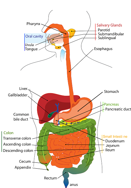
To reach the colon and to be able to specifically deliver and absorb the drug there, the dosage forms must be formulated taking into account the obstacles of the gastrointestinal tract. The various strategies developed to achieve this goal have used the specific characteristics of this organ, i.e. transit time, pH, microflora, enzymes, disease and the colonic environment. Nevertheless, these parameters can vary from one individual to the next and also according to the pathological condition and diet.


Physiological Factors

Gastrointestinal transit

Gastrointestinal transit time is important for nearly all orally targeting delivery systems. The drug delivery systems first enter in to stomach and small intestine via mouth and then reach colon. In fasted state, the motility proceeds through four phases occurring in stomach and small intestine that span over a period of 2-3 h. Phase I is a quiescent period of 40-60 min, Phase II consists of intermittent contractions for a period of 40-60 min. Phase III is a period of intense contractions sweeping material out of the stomach and down the small intestine followed by Phase IV with contractions dissipating. The feeding state affects the normal pattern by irregular contractile activity.  

It has been well documented that gastric emptying varies with different types of dosage forms. Examples of gastric residence times of single-unit tablets are given in Table 1 (Abrahamsson, 1993). It has been generally accepted that liquid emptying follows a monoexponential process and digestible solids empty in a linear fashion with time.


Small intestinal transit

Normally, transit times through the small intestine generally found to be 3-4 h. Liquids, small solids (beads, small tablets), and larger capsule-sized units moved essentially at the same rates and the transit is unaffected by food status (Davis, 1986). In a more recent study concerning dosing in relation to the timing of food intake, found that although SIT is relatively independent of food and dosage form, it was actually shortened significantly if the dose is given 30 min before food intake. This can have adverse impact on the in vivo performance of the dosage forms.


Colonic Transit

In the stomach and small intestine, food residue and endogenous secretions are exposed to an essentially sterile environment through which their transit can be measured by hours. On entering the large intestine, dosage forms encounter a rich bacterial flora and transit through the large intestine can be as long as several days. It was reported that overall mean transit time is 36 h with a range of 1 to 72 h and that the transit of liquids and small solids is equal (Phillips., 1993). Thus, absorption from colon may be incomplete and erratic depending on the dose and physicochemical properties of a particular drug. In general, absorption of an insoluble drug with high dose or a drug with limited permeability is unfavorable in this region because of the limited volume of fluid available for dissolution and the significantly reduced surface area.

 Pharmacology Study Notes Colon COLON SPECIFIC DRUG DELIVERY SYSTEM pdf Pharmacology Study Notes Colon COLON SPECIFIC DRUG DELIVERY SYSTEM Pharmacology Study Notes Colon COLON SPECIFIC DRUG DELIVERY SYSTEM

Table 1. Gastrointestinal transit times for felodipine CR hydrophilic matrix

section Gastric emptying (h) Small intestine transit (h) Colon arrival (h)
Fasting Fed Fasting Fed Fasting Fed
Mean 0.6 3.2 4.7 5.1 5.3 8.3
Range 0.1-1.1 1.9-4.8 3.9-5.9 2.2-7.7 4.0-7.0 6.0-11.0
P <0.001 >0.05 >0.01


pH in the Colon

The pH gradient in the GIT is not in an increased order and is subjected to both inter- and intra-subject variations. In stomach the pH is 1.5 – 2.0 and 2 – 6 in fasted and fed conditions, respectively. The acidic pH is responsible for the degradation of various pH sensitive drugs and enteric coating may prevent it. In small intestine, the pH increases slightly from 6.6 – 7.5. On entry into the colon, the pH dropped to 6.4 in right colon. The pH of mid colon was found to be 6.6 and in the left colon, 7.0 (Evans et al., 1988).

Colonic pH has been shown reduced in disease state. The mean pH in a group of 7 patients with untreated ulcerative colitis was 4.7 whereas in 5 patients receiving treatment it was 5.5 (Raimundo et al., 1992).

Colonic microflora

The human colon is a dynamic and ecologically diverse environment, containing over 400 distinct species of bacteria with a population of 1011 to 1012 CFU/mL (Cummings et al., 1991), with Bacteroides, Bifidobacterium, Eubacterium, Lactobacillus, etc greatly outnumbering other species. For example, it was reported that Bacteroides, Bifidobacterium and Eubacterium could constitute as much as over 60% of the total cultivable flora (Salyers, 1984). These bacteria produce a wide spectrum of enzymes that, being reductive and hydrolytic in nature, are actively involved in many processes in the colon, such as carbohydrate and protein fermentation, bile acid and steroid transformation, metabolism of xenobiotic substances, as well as the activation and destruction of potential mutagenic metabolites. Nitroreductase, azoreductase, N-oxide and sulfoxide reductase are the most extensively investigated reductive enzymes, while glucosidase and glucuronidase are the most extensively studied hydrolytic enzymes. The primary source of nutrition for these anaerobic bacteria is carbohydrates such as non-starch polysaccharides (i.e., dietary fibers) from the intestinal chime. It is well established that non-starch polysaccharides are fermented during transit through the colon and the breakdown in the stomach and small intestine is negligible. Enzymes responsible for the degradation of polysaccharides include α-L-arabinofuranosidase, β-D-fucosidase,  β-D-

galactosidase, β-Dglucosidase, β-xylosidase, with the last three enzymes being the most active (Englyst et al., 1987). Additionally, the composition of colonic bacteria and corresponding enzymes can be influenced by many factors, including age, diet, diseases, medication such as antibiotics, and geographic regions (Mueller et al., 2006). A unique feature of colon microflora is that the growth and activity of certain specific species, most notably bifidobacteria and lactobacilli, can be selectively stimulated by nondigestible oligosaccharides which are known as prebiotics. Similar bacteriological data were observed in the rats fed with indigestible oligosaccharides where the caecal bifidobacteria population was higher than in the controls (Campbell et al., 1997).

Volume of the ascending colon

Up to 1,500 g of liquids and undigested materials (dietary fibers, resistant starch, partially degraded polysaccharides proteins, mucins, exfoliated epithelial cells, etc.) enters colon per day, which act as the substrates for microflora fermentation. Water together with the products of the fermentation and other nutrients was efficiently absorbed in the colon, condensing the contents into feces through the transit in the colon for eventual defecation. Therefore, it is very likely that the ascending colon contains the largest quantity of liquid. It would be expected that the low water–high gas environment of the transverse colon limits dissolution of materials.  The moisture content of caecal contents is believed to be about 86% (Cummings and Macfarlane, 1991). The volume of the ascending colon was measured in healthy subjects using a single photon emission computed tomography (SPECT) by acquiring the imaging of the ascending colon filled with 99Tcm-labelled Amberlite pellets, and was found to be 170±40 ml (Badley., 1993). If the moisture content in the ascending colon is approximately comparable to that of caecal contents, the quantity of fluid in the ascending colon should be 146±34 ml.

[DOC Pdf PPT] Physiology

Pharmaceutics notes B Pharmacy M pharmacy Study Material

Disease and the Colonic Environment

General intestinal diseases such as inflammatory bowel disease, Crohn’s disease, constipation and diarrhea may affect the release and absorption of colon specific drug delivery systems. All the specific approaches so far mentioned rely on the concept that enzymes produced by colonic microflora provide the trigger for specific delivery of fermentable coatings, anti-inflammatory azobond drugs, and other prodrugs to the cecum. Carrette and co-workers (1995) demonstrated that in patients with active Crohn’s disease, the metabolic activity of digestive flora (assessed on the activity of fecal glycosidases) was decreased. Azoreductase activity in feces of 14 patients with active Crohn’s disease was 20% of that of healthy subjects and similarly, beta-D-glucosidase and beta-D-glucuronidase activities in fecal homogenates incubated under anaerobic conditions were also decreased in patients. These data probably reflect large-bowel hypermotility and the associated diarrhea, leading to lower bacterial mass in the colon and might contribute to the therapeutic failure of targeting mechanisms in active ileocolic and colic Crohn’s disease.