Pharmacology Notes: PPT PDF – ANTICANCER DRUGS – What is Cancer? Types/ Causes

Pharmacology Notes PPT PDF - ANTICANCER DRUGS - What is Cancer Types Causes

Pharmacology Notes

ANTICANCER DRUGS

Cancer cells have lost the normal regulatory mechanisms that control cell growth and multiplication.

What is Cancer?

• Cancer cell have lost their ability to differentiate (that means to specialize). Cancer refers to any one of a large number of diseases characterized by the development of abnormal cells that divide uncontrollably and have the ability to infiltrate and destroy normal body tissue. Cancer often has the ability to spread throughout your body.

Types of Cancer?

• Benign cancer cell stay at the same place
Malignant cancer cells invade new tissues to set up secondary tumors, a process known as metastasis

Causes of cancer

Common Causes of Cancer:

Smoking and Tobacco. Diet and Physical Activity. Sun and Other Types of Radiation. Viruses and Other Infections

• Chemicals causing cancer are called mutagens
• Cancer can be caused by chemicals, life style (smoking), and viruses

Gene mutations

A gene mutation can instruct a healthy cell to Allow rapid growth or Fail to stop uncontrolled cell growth or cells lose the controls (tumor suppressor genes) or even Make mistakes when repairing DNA errors

Definitions of cancer

genes that are related to cause cancer are called oncogenes.
Genes that become onogenic upon mutation are called protooncogenes.

Pharmacology Notes PPT PDF - ANTICANCER DRUGS - What is Cancer Types Causes

General signs and symptoms of cancer

Unexplained weight loss
Fever
Fatigue
Pain
Skin changes
Darker looking skin (hyperpigmentation)
Yellowish skin and eyes (jaundice)
Reddened skin (erythema)
Itching (pruritis)
Excessive hair growth
Change in bowel habits or bladder function
Long-term constipation, diarrhea,
Sores that do not heal
White patches inside the mouth or white spots on the tongue
Unusual bleeding or discharge
Thickening or lump in the breast or other parts of the body
Indigestion or trouble swallowing
Recent change in a wart or mole or any new skin change
Nagging cough or hoarseness

Top 10 Anti Cancer Drugs

anti cancer drugs list ppt pharmacology

List of Anti cancer Drugs

ALKYLATING AGENTS:

BUSULFAN
CARMUSTINE (BCNU)
CYCLOPHOSPHAMIDE
DACARBAZINE
LOMUSTINE (CCNU)
MECHLORETHAMINE
MELPHALAN
THIOTEPA

NATURAL PRODUCTS

BLEOMYCIN
DACTINOMYCIN
DAUNORUBICIN
DOXORUBICIN
ETOPOSIDE (VP-16)
IRINOTECAN
MITOMYCIN C
PACLITAXEL
VINBLASTINE
VINCRISTINE

MISCELLANEOUS:

Angiostatin
AMSACRINE
L-asparaginase
Bortezomib
CARBOPLATIN
CISPLATIN
Erlotinib
Gefitinib
Hydroxyurea
Imatinib
Pentostatin
PROCARBAZINE
Thalidomide

ANTIMETABOLITES:

Azathioprine
5-fluorouracil
6-thioguanine
6-mecaptopurine
Cytarabine (ara-c)
Gemcitabine
Methotrexate

IMMUNOTHERAPY:

Alemtuzumab
Aminoglutethimide
Bevacizumab
Cetuximab
Cyclosporine
Dexamethasone
Edrecolomab
Gemtuzumab
Ibritumomab
Interferon α
Interleukin 2
Interleukin-12
Prednisone
Rituximab
Tacrolimus (fk506)
Tositumomab
Trastuzumab
Tumour necrosis factor α

HORMONES and RELATED AGENTS:

Aminoglutethimide
Anastrozole
Exemestane
Flutamide
Letrozole
Goserelin
Leuprolide
Letrozole
Tamoxifen

SUPPORTING AGENTS:

Allopurinol
Erythropoietin
Filgrastim
Interleukin 11
Leucovorin
MESNA
Sargramostim (GM-CSF)

anti cancer drugs ppt pdf notes b pharm m pharm medicos d pharm pharmacology

Pharmacology anti cancer drugs ppt pdf notes b pharm m pharm medicos d pharm

anti neoplastic anti cancer drugs ppt pdf notes b pharm m pharm medicos d pharm

Anticancer drugs pharmacology pdf anticancer drugs list pdf classification of anticancer drugs wikipedia anticancer drugs classification ppt classification of anticancer drugs with mechanism of action classification of anticancer agents anticancer drugs classification mnemonics top 10 anti cancer drugs.

Homology Modelling of Protein Steps Tools Software Tutorial PDF PPT Papers

Homology Modelling of Protein Steps Tools Software Tutorial PDF PPT Papers

What is Homology Modelling?

Homology modelling allows users to safely use rapidly generated in silico protein models in all the contexts where today only experimental structures provide a solid basis: structure-based drug design, analysis of protein function, interactions, antigenic behavior, and rational design of proteins with increased stability or novel functions. In addition, protein modeling is the only way to obtain structural information if experimental techniques fail. Many proteins are simply too large for NMR analysis and cannot be crystallized for X-ray diffraction.

Homology Modelling of Protein Steps Tools Software Tutorial PDF PPT Papers

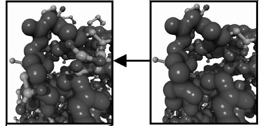
Among the major approaches to three-dimensional (3D) structure prediction, homology modeling is the easiest one.
In the Homology Modelling, structure of a protein is uniquely determined by its amino acid sequence (Epstain, Goldberger, and Anfinsen, 1963). Knowing the sequence should, at least in theory, suffice to obtain the structure.
2. During evolution, the structure is more stable and changes much slower than the associated sequence, so that similar sequences adopt practically identical structures, and distantly related sequences still fold into similar structures. This relationship was first identified by Chothia and Lesk (1986) and later quantified by Sander and Schneider (1991). Thanks to the exponential growth of the Protein Data Bank (PDB), Rost (1999) could recently derive a precise limit for this rule. As long as the length of two sequences and the percentage of identical residues fall in the region marked as “safe,” the two sequences are practically guaranteed to adopt a similar structure.

Homology Modelling or Protein Modelling Example

Imagine that we want to know the structure of sequence A (150 amino acids long,). We compare sequence A to all the sequences of known structures stored in the PDB (using, for example, BLAST), and luckily find a sequence B (300 amino acids long) containing a region of 150 amino acids that match sequence A with 50% identical residues. As this match (alignment) clearly falls in the safe zone (Fig. 25.1), we can simply take the known structure of sequence B
(the template), cut out the fragment corresponding to the aligned region, mutate those amino acids that differ between sequences A and B, and finally arrive at our model for structure A. Structure A is called the target and is of course not known at the time of modeling.

Homology Modelling of Protein Steps Tools Software Tutorial PDF PPT

Homology Modelling Steps

In practice, homology modeling is a multistep process that can be summarized in seven steps:
1. Template recognition and initial alignment
2. Alignment correction
3. Backbone generation
4. Loop modeling
5. Side-chain modeling
6. Model optimization
7. Model validation

At almost all the steps choices have to be made. The modeler can never be sure to make the best ones, and thus a large part of the modeling process consists of serious thought about how to gamble between multiple seemingly similar choices. A lot of research has been spent on teaching the computer how to make these decisions, so that homology models can be built fully automatically. Currently, this allows modelers to construct models for about 25% of the amino acids in a genome, thereby supplementing the efforts of structural genomics projects.

Homology_Modelling – Protein PPT

homology modeling

Protein Homology modelling steps ppt Structures

Homology Modelling Steps, Homology Modelling Software, Homology Modelling Ppt, Homology Modelling Pdf, Homology Modeling Server, Protein Modelling Bioinformatics, Homology Modeling Tutorial, Homology Modelling Slideshare

ANATOMY & PHYSIOLOGY Of Human Respiratory Tract

ANATOMY & PHYSIOLOGY Of Human Respiratory Tract

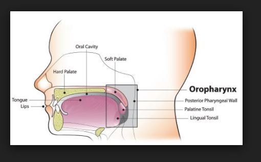
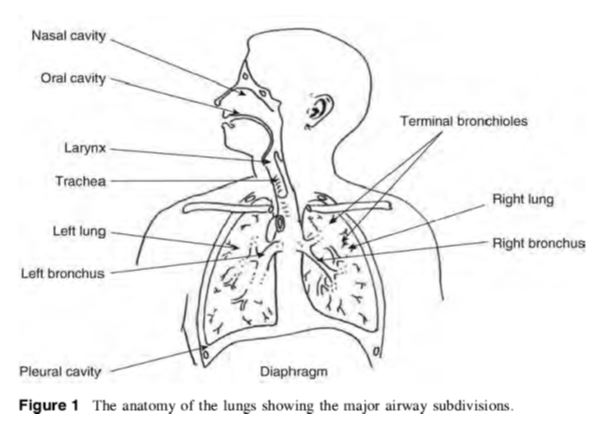
Anatomy and Physiology Of human respiratory system is a complicated organ system of very close structure– function relationships. The system consisted of two regions: the conducting airway and the respiratory region. The airway is further divided into many folds: nasal cavity and the associated sinuses, and the nasopharynx, oropharynx, larynx, trachea, bronchi, and bronchioles. The respiratory region consists of respiratory bronchioles, alveolar ducts, and alveolar sacs.

ANATOMY AND PHYSIOLOGY OF HUMAN RESPIRATORY TRACT:

               The respiratory system works with the circulatory system to deliver oxygen from the lungs to the cells and remove carbon dioxide, and return it to the lungs to be exhaled. The exchange of oxygen and carbon dioxide between the air, blood and body tissues is known as respiration. Healthy lungs take in about 1 pint of air about 12–15 times each minute. All of the blood in the body is passed through the lungs every minute. The respiratory tract is divided into two main parts: the upper respiratory tract, consisting of the nose, nasal cavity and the pharynx; and the lower respiratory tract consisting of the larynx, trachea, bronchi and the lungs The trachea, which begins at the edge of the larynx, divides into two bronchi and continues into the lungs. The trachea allows air to pass from the larynx to the bronchi and then to the lungs. The bronchi divide into smaller bronchioles which branch in the lungs forming passageways for air. The terminal parts of the bronchi are the alveoli. The alveoli are the functional units of the lungs and they form the site of gaseous exchange     

ANATOMY & PHYSIOLOGY Of Human Respiratory Tract

            The blood barrier between the alveolar space and the pulmonary capillaries is very thin to allow for rapid gas exchange. During inspiration, oxygen diffuses through the alveoli walls and the interstitial space, into the blood. Carbon dioxide diffuses in the opposite direction during exhalation. Alveoli are small and there are approximately 300 million of them in each lung. Although alveoli are tiny structures, they have a very large surface area in total (~100 m2) for performing efficient gas exchange.

                     The alveoli form a honeycomb of cells around the spiral, cylindrical surface of the alveolar duct. The exposed alveolar surface is normally covered with a surface film of lipoprotein material.

                      There are several types of pulmonary alveolar cells. Type I (or small type A), are non-phagocytic, membranous pneumocytes. These surface-lining epithelial cells are approximately 5 μm in thickness and possess thin squamous cytoplasmic extensions that originate from a central nucleated portion. These portions do not have any organelles and hence they are metabolically dependent on the central portion of the cell. This reduces their ability to repair themselves if damaged. Attached to the basement membrane are the larger alveolar cells (Type II, type B or septal cells). These rounded, granular, epithelial pneumocytes are approximately 10 to 15 μm tick. There are 6 to 7 cells per alveolus and these cells possess great metabolic activity. They are believed to produce the surfactant material that lines the lung and to be essential for alveolar repair after damage from viruses or chemical agents.

             Amongst, the important roles of the lungs, one can cite: (i) supply oxygen, (ii) remove wastes and toxins, and (iii) defend against hostile intruders. The lungs have three dozen distinct types of cells. Some of these cells scavenge foreign matter. Others have cilia that sweep the mucous membranes lining the smallest air passages. Some cells act on blood pressure control, while others spot infection invaders.

 anatomy of lungs            

      The respiratory system is susceptible to a number of diseases, and the lungs are prone to a wide range of disorders caused by genetic factors, infection and pollutants in the air. The most common problems of the respiratory system are:

  • Asthma
  • Bronchiolitis
  • Chronic obstructive pulmonary disease (COPD)
  • Common cold
  • Cough
  • Cystic fibrosis (CF)
  • Lung cancer
  • Pneumonia
  • Pulmonary hypertension

PRINCIPAL MECHANISMS OF RESPIRATORY DEPOSITION

            The deposition of inhaled particles in the different regions of the respiratory system is very complex, and depends on many factors. Some of the factors influencing respiratory deposition include:

  • Breathing rate
  • Mouth or nose breathing
  • Lung volume
  • Respiration volume
  • Health of the individual
  • Bifurcations in the airways result in a constantly changing hydrodynamic flow field.

Depending on the particle size, airflow, and location in the respiratory system, particle deposition occurs via on of the following principal mechanisms:

Impaction

         Each time the airflow changes due to a bifurcation in the airways, the suspended particles tend to travel along their original path due to inertia and may impact on an airway surface. This mechanism is highly dependent on aerodynamic diameter, since the stopping distance for very small particles is quite low. Impaction occurs mostly in the case of larger particles that are very close to airway walls, near the first airway bifurcations. Therefore, deposition by impaction is greatest in the bronchial region. Impaction accounts for the majority of particle deposition on a mass basis.

 Sedimentation

           Sedimentation is the settling out of particles in the smaller airways of the bronchioles and alveoli, where the air flow is low and airway dimensions are small. The rate of sedimentation is dependent on the terminal settling velocity of the particles, so sedimentation plays a greater role in the deposition of particles with larger aerodynamic diameters. Hygroscopic particles may grow in size as they pass through the warm, humid air passages, thus increasing the probability of deposition by sedimentation.

 Interception

            Interception occurs when a particle contacts an airway surface due to its physical size or shape. Unlike impaction, particles that are deposited by interception do not deviate from their air streamlines. Interception is most likely to occur in small airways or when the air streamline is close to an airway wall. Interception is most significant for fibers, which easily contact airway surfaces do to their length. Furthermore, fibers have small aerodynamic diameters relative to their size, so they can often reach the smallest airways.


Diffusion

Diffusion is the primary mechanism of deposition for particles less than 0.5 microns in diameter and is governed by geometric rather than aerodynamic size. Diffusion is the net transport of particles from a region of high concentration to a region of lower concentration due to Brownian motion. Brownian motion is the random wiggling motion of a particle due to the constant bombardment of air molecules. Diffusional deposition occurs mostly when the particles have just entered the nasopharynx, and is also most likely to occur in the smaller airways of the pulmonary (alveolar) region, where air flow is low.


 Absorption – bioavailability of drugs

Although inhaled drugs have been used for over 50 years to treat airway disease and are in development or being considered for the treatment of many other lung diseases, insulin is at present time the only one representative inhaled drug on the market for systemic disease. Exubera® (insulin human [rDNA origin] inhalation powder is the first diabetes treatment which can be inhaled. Exubera® helps control high blood sugar, works in adults with type 1 diabetes and with type 2 diabetes as well This therapeutic success has lead a number of other companies to investigate and to advance clinical trials as inhaled formulations for systemic applications with a variety of large molecules (leuprolide, a luteinizing hormone-releasing hormone (LHRH) analogue, …). Recent advances in the development of particle technologies and devices now make it possible to formulate, stabilize, and accurately deliver almost any drug to the lungs.

             The pulmonary membrane is naturally permeable to small molecule drugs and to many therapeutic peptides and proteins. The epithelium of the lung, the significant barrier to absorption of inhaled drugs, is thick (50–60 μm) in the trachea, but diminishes in thickness to an extremely thin 0.2 μm in the alveoli. The change in cell types and morphology going from trachea, bronchi, and bronchioles to alveoli is very dramatic. The lungs are for more permeable to macromolecules than any other portal of entry into the body. Some of the most promising therapeutic agents are peptides and proteins, which could be inhaled instead of injected, thereby improving compliance .Particularly, peptides that have been chemically altered to inhibit peptidase enzymes exhibit very high bioavailabilities by the pulmonary route .Indeed, natural mammalian peptides, les than 30 amino acids (somatostatin, vaso active intestinal peptide [VIP], and glucagons), are broken down in the lung by ubiquitous peptidases and have very poor bioavailabilities. Conversely, proteins with molecular weights between 6000 and 50,000 Da are relatively resistant to most peptidases and have good bioavailabilities following inhalation. For larger proteins, the bioavailabilities and absorption mechanisms are not well completely elucidated.

ADVANTAGES OF PULMONARY DRUG DELIVERY SYSTEM


  1. The ability to nebulize viscous drug formulations for pulmonary delivery, thereby overcoming drug solubility issues with the ability to use lipid, water or lipid/water emulsions as drug carriers.
  2. Ability to nebulize viscous liquids into droplets in the 2-5μm range regardless of the carrier composition solubility which would allow for a wide range of drug formulation options.
  3. Increased drug delivery efficacy due to size-stable aerosol droplets with reduced

hygroscopic growth and evaporative shrinkage.

  1. Liposomal drug formulations remain stable when nebulized.
  2. Ability to nebulize protein-containing solutions.
  3. For hand held inhaler applications, drug does not need to be emulsified in liquefied nebulizing gas to achieve aerosolization.

Colon targeting – Colonic drug delivery Uses Pharmacology Study Material – Project Thesis Title

colon targeting Colonic drug delivery Uses Pharmacology Study Material Project Thesis Title

Colon targeting – Colonic drug delivery Uses:

Pharmacology Study Material – Project Thesis Title is here to help students research scholars to have a brief study on colonic drug delivery system advantages

Numerous drug entities based on oral delivery have been successfully commercialized, but many others are not readily available by oral administration, which are incompatible with the physical and/or chemical environments of the upper gastrointestinal tract (GIT) and/or demonstrate poor uptake in the upper GI tract. Due to the lack of digestive enzymes, colon is considered as suitable site for the absorption of various drugs. Over the past two decades the major challenge for scientist is to target the drugs specifically to the colonic region of GIT. Previously colon was considered as an innocuous organ solely responsible for absorption of water, electrolytes & temporary storage of stools. But now it is accepted as important site for drug delivery.

DOWNLOAD PDF pharmacology notes

Colon targeting – Colonic drug delivery Uses Pharmacology Study Material – Project Thesis Title Pdf doc Colon targeting – Colonic drug delivery Uses Pharmacology Study Material – Project Thesis Title  colon targeting Colonic drug delivery Uses Pharmacology Study Material Project Thesis Title

Colon targeting is used to treat:-

  • Seriousness from constipation & diarrhea to the debilitating inflammatory bowel diseases (Ulcerative colitis & Crohn’s disease) through to colon carcinoma which is two third cause of cancer in both man & women.
  • Colon can be utilized as portal for the entry of drugs into the blood stream for the systemic therapy.
  • Colon having the lower level of luminal & mucosal digestive enzymes as compared with the small intestine reduces the chances of drug degradation. e.g., to facilitate absorption of acid and enzymatically labile materials, especially proteins and peptides (Ikesue et al., 1991).
  • Colon delivery also a mean of achieving chronotherapy of disease that is sensitive to circadian rhythm such as asthma & arthritis (Quadros et al., 1995).
  • Targeted delivery ensures the direct treatment at the disease site, lower dosing, & reduction in side effects.
  • Colonic drug delivery is also found useful for improving systemic absorption of drugs like nitrendipine, metoprolol, theophylline, isosorbide mononitrate etc.
  • ANOTOMY & PHYSIOLOGY OF COLON Pharmacology study material for pharmacy

Pharmacology Study Material – Project Thesis Title

 The rectal route has traditionally been used to administer medicaments in the form of suppositories and enemas to the distal gut, although such formulations rarely succeed in spreading beyond the descending colon. Also, the rectal route is not convenient or acceptable for most patients and hence the oral route is the preferred route of drug administration. However, colonic drug delivery via the oral route is not without its challenges. The colon constitutes the most distal segment of the gastrointestinal tract and so an orally administered formulation must retard drug release in the upper gastrointestinal regions but release the drug promptly on entry into the colon.

  Retardation of drug release in the diverse and hostile conditions of the stomach and small intestine is not easily achieved, since the dosage form will be subjected to a physical and chemical assault that is designed to break down ingested materials. While in the colon, the low fluid environment and viscous nature of luminal contents may hinder the dissolution and release of the drug from the formulation. Moreover, the resident colonic microflora may impact on the stability of the released drug via metabolic degradation. In spite of these potential difficulties, a variety of approaches have been used and systems have been developed for the purpose of achieving colonic targeting. Targeted drug delivery is reliant on the identification and exploitation of a characteristic that is specific to the target organ. In the context of colonic targeting, the exploitable gastrointestinal features include pH, transit time, pressure, bacteria and prodrug approach.

DRUGS SUITABLE FOR COLONIC DRUG DELIVERY


[PPT PDF] Pharmaceutical Water System Validation – IDENTIFICATION OF MICROORGANISMS

[PPT PDF] Pharmaceutical Water System Validation - IDENTIFICATION OF MICROORGANISMS

IDENTIFICATION OF MICROORGANISMS – Pharmaceutical Water System Validation

Identifying the isolates recovered from water monitoring methods may be important in instances where specific waterborne microorganisms may be detrimental to the products or processes in which the water is used. Microorganism information such as this may also be useful when identifying the source of microbial contamination in a product or process. Often a limited group of microorganisms is routinely recovered from a water system. After repeated recovery and characterization, an experienced microbiologist may become proficient at their identification based on only a few recognizable traits such as colonial morphology and staining characteristics. This may allow for a reduction in the number of identifications to representative colony types, or, with proper analyst qualification, may even allow testing short cuts to be taken for these microbial identifications.

ALERT AND ACTION LEVELS AND SPECIFICATIONS

Though the use of alert and action levels is most often associated with microbial data, they can be associated with any attribute. In pharmaceutical water systems, almost every quality attribute, other than microbial quality, can be very rapidly determined with near-real time results. These short-delay data can give immediate system performance feedback, serving as ongoing process control indicators. However, because some attributes may not continuously be monitored or have a long delay in data availability (like microbial monitoring data), properly established Alert and Action Levels can serve as an early warning or indication of a potentially approaching quality shift occurring between or at the next periodic monitoring. In a validated water system, process controls should yield relatively constant and more than adequate values for these monitored attributes such that their Alert and Action Levels are infrequently broached.

As process control indicators, alert and action levels are designed to allow remedial action to occur that will prevent a system from deviating completely out of control and producing water unfit for its intended use. This “intended use” minimum quality is sometimes referred to as a “specification” or “limit”. In the opening paragraphs of this chapter, rationale was presented for no microbial specifications being included within the body of the bulk water (Purified Water and Water for Injection) monographs. This does not mean that the user should not have microbial specifications for these waters. To the contrary, in most situations such specifications should be established by the user. The microbial specification should reflect the maximum microbial level at which the water is still fit for use without compromising the quality needs of the process or product where the water is used. Because water from a given system may have many uses, the most stringent of these uses should be used to establish this specification.

Where appropriate, a microbial specification could be qualitative as well as quantitative. In other words, the number of total microorganisms may be as important as the number of a specific microorganism or even the absence of a specific microorganism. Microorganisms that are known to be problematic could include opportunistic or overt pathogens, nonpathogenic indicators of potentially undetected pathogens, or microorganisms known to compromise a process or product, such as by being resistant to a preservative or able to proliferate in or degrade a product. These microorganisms comprise an often ill-defined group referred to as “objectionable microorganisms”. Because objectionable is a term relative to the water’s use, the list of microorganisms in such a group should be tailored to those species with the potential to be present and problematic. Their negative impact is most often demonstrated when they are present in high numbers, but depending on the species, an allowable level may exist, below which they may not be considered objectionable.

[PPT PDF] Pharmaceutical Water System Validation – IDENTIFICATION OF MICROORGANISMS IDENTIFICATION OF MICROORGANISMS – Pharmaceutical warer system ppt [PPT PDF] Pharmaceutical Water System Validation - IDENTIFICATION OF MICROORGANISMS

As stated above, alert and action levels for a given process control attribute are used to help maintain system control and avoid exceeding the pass/fail specification for that attribute. Alert and action levels may be both quantitative and qualitative. They may involve levels of total microbial counts or recoveries of specific microorganisms. Alert levels are events or levels that, when they occur or are exceeded, indicate that a process may have drifted from its normal operating condition. Alert level excursions constitute a warning and do not necessarily require a corrective action. However, alert level excursions usually lead to the alerting of personnel involved in water system operation as well as QA. Alert level excursions may also lead to additional monitoring with more intense scrutiny of resulting and neighboring data as well as other process indicators. Action levels are events or higher levels that, when they occur or are exceeded, indicate that a process is probably drifting from its normal operating range. Examples of kinds of action level “events” include exceeding alert levels repeatedly; or in multiple simultaneous locations, a single occurrence of exceeding a higher microbial level; or the individual or repeated recovery of specific objectionable microorganisms. Exceeding an action level should lead to immediate notification of both QA and personnel involved in water system operations so that corrective actions can immediately be taken to bring the process back into its normal operating range. Such remedial actions should also include efforts to understand and eliminate or at least reduce the incidence of a future occurrence. A root cause investigation may be necessary to devise an effective preventative action strategy. Depending on the nature of the action level excursion, it may also be necessary to evaluate its impact on the water uses during that time. Impact evaluations may include delineation of affected batches and additional or more extensive product testing. It may also involve experimental product challenges.

Alert and action levels should be derived from an evaluation of historic monitoring data called a trend analysis. Other guidelines on approaches that may be used, ranging from “inspectional”to statistical evaluation of the historical data have been published. The ultimate goal is to understand the normal variability of the data during what is considered a typical operational period. Then, trigger points or levels can be established that will signal when future data may be approaching (alert level) or exceeding (action level) the boundaries of that “normal variability”. Such alert and action levels are based on the control capability of the system as it was being maintained and controlled during that historic period of typical control.

In new water systems where there is very limited or no historic data from which to derive data trends, it is common to simply establish initial alert and action levels based on a combination of equipment design capabilities but below the process and product specifications where water is used. It is also common, especially for ambient water systems, to microbiologically “mature” over the first year of use. By the end of this period, a relatively steady state microbial population (microorganism types and levels) will have been allowed or promoted to develop as a result of the collective effects of routine system maintenance and operation, including the frequency of unit operation rebeddings, backwashings, regenerations, and sanitizations. This microbial population will typically be higher than was seen when the water system was new, so it should be expected that the data trends (and the resulting alert and action levels) will increase over this “maturation” period and eventually level off.

Pharmaceutical water System

A water system should be designed so that performance-based alert and action levels are well below water specifications. With poorly designed or maintained water systems, the system owner may find that initial new system microbial levels were acceptable for the water uses and specifications, but the mature levels are not. This is a serious situation, which if not correctable with more frequent system maintenance and sanitization, may require expensive water system renovation or even replacement. Therefore, it cannot be overemphasized that water systems should be designed for ease of microbial control, so that when monitored against alert and action levels, and maintained accordingly, the water continuously meets all applicable specifications.

An action level should not be established at a level equivalent to the specification. This leaves no room for remedial system maintenance that could avoid a specification excursion. Exceeding a specification is a far more serious event than an action level excursion. A specification excursion may trigger an extensive finished product impact investigation, substantial remedial actions within the water system that may include a complete shutdown, and possibly even product rejection.

Another scenario to be avoided is the establishment of an arbitrarily high and usually nonperformance based action level. Such unrealistic action levels deprive users of meaningful indicator values that could trigger remedial system maintenance. Unrealistically high action levels allow systems to grow well out of control before action is taken, when their intent should be to catch a system imbalance before it goes wildly out of control.

Because alert and action levels should be based on actual system performance, and the system performance data are generated by a given test method, it follows that those alert and action levels should be valid only for test results generated by the same test method. It is invalid to apply alert and action level criteria to test results generated by a different test method. The two test methods may not equivalently recover microorganisms from the same water samples. Similarly invalid is the use of trend data to derive alert and action levels for one water system, but applying those alert and action levels to a different water system. Alert and action levels are water system and test method specific.

Nevertheless, there are certain maximum microbial levels above which action levels should never be established. Water systems with these levels should unarguably be considered out of control. Using the microbial enumeration methodologies suggested above, generally considered maximum action levels are 100 cfu per mL for Purified Water and 10 cfu per 100 mL for Water for Injection. However, if a given water system controls microorganisms much more tightly than these levels, appropriate alert and action levels should be established from these tighter control levels so that they can truly indicate when water systems may be starting to trend out of control. These in-process microbial control parameters should be established well below the user-defined microbial specifications that delineate the water’s fitness for use.

Special consideration is needed for establishing maximum microbial action levels for Drinking Water because the water is often delivered to the facility in a condition over which the user has little control. High microbial levels in Drinking Water may be indicative of a municipal water system upset, broken water main, or inadequate disinfection, and therefore, potential contamination with objectionable microorganisms. Using the suggested microbial enumeration methodology, a reasonable maximum action level for Drinking Water is 500 cfu per mL. Considering the potential concern for objectionable microorganisms raised by such high microbial levels in the feedwater, informing the municipality of the problem so they may begin corrective actions should be an immediate first step. In-house remedial actions may or may not also be needed, but could include performing additional coliform testing on the incoming water and pretreating the water with either additional chlorination or UV light irradiation or filtration or a combination of approaches.

Source : USP

Expert Committee : (PW05) Pharmaceutical Waters 05

USP29–NF24 Page 3056

Pharmacopeial Forum : Volume No. 30(5) Page 1744

Pharmaceutical Water System Ppt,

Pharmaceutical Water Systems,

Purified Water Specification As Per Usp,

Pharmaceutical Water System Design Operation And Validation Pdf,

pharmaceutical water system design operation and validation,

pharmaceutical water system ppt – What is Pharmaceutical water,

purified water & Water for Injection SOP as per usp,

Pharmaceutical Water Systems: Storage & Distribution Systems,

pharmaceutical water system : Inspection of Pharmaceutical water systems,

pharmaceutical water Production : Water purification systems,

Pharmaceutical Water Systems: Types: Water quality specifications,

Pharmaceutical Water System: principles for pharmaceutical water systems

M Pharmacy Parmaceutics Project : APPROACHES TO COLON-SPECIFIC DRUG DELIVERY [Ceutics]

M Pharmacy Parmaceutics Project APPROACHES TO COLON-SPECIFIC DRUG DELIVERY [Ceutics]

M Pharmacy Project : APPROACHES TO COLON-SPECIFIC DRUG DELIVERY

APPROACHES TO COLON-SPECIFIC DRUG DELIVERY

In recent years, a large number of solid formulations targeting the lower parts of the GI tract, especially the colon, have been reported. These formulations may be broadly divided into four types, which are

  1. pH-dependent system designed to release a drug in response to change in pH,
  2. Time controlled ( or Time-dependent) system designed to release a drug after a predetermined time,
  3. Microbially-controlled system making use of the abundant enterobacteria in the colon,
  4. Enzyme-based systemsProdrug, and
  5. Pressure-dependent system making use of luminal pressure of the colon.

Among these, first three are most widespread formulation technologies being developed for pharmaceutical market.


pH-DEPENDENT SYSTEMS

Solid formulations for colonic delivery that are based on pH-dependent drug release mechanism are similar to conventional enteric-coated formulations but they differ in target site for delivery and therefore type of enteric polymers. In contrast to conventional enteric-coated formulations, colonic formulations are designed to deliver drugs to the distal (terminal) ileum and colon, and utilize enteric polymers that have relatively higher threshold pH for dissolution (Dew et al., 1982; Tuleu et al., 2001). Most commonly used polymers (Table 2) are derivatives of acrylic acid and cellulose. These polymers have ability to withstand an environment ranging from low pH (~1.2) to neutral pH (~7.5) for several hours. Apparently, it is highly desirable for pH-dependent colonic formulations to maintain their physical and chemical integrity during passage through the stomach and small intestine and reach the large intestine where the coat should disintegrate to release the drug locally. It should be however noted that GI fluids might pass through the coat while the dosage form transits through the small intestine. This could lead to premature drug release in the upper parts of GI tract and as a result loss of therapeutic efficacy may occur. One approach to overcome this problem is to apply

higher coating levels of enteric polymers; however, this also allows influx of GI fluids through the coat, and the thicker coats often rupture under the influence of contractile activity in the stomach. In general, the amount of coating required depends upon the solubility characteristics (solubility, dose/solubility ratio) of the drug, desired release profile and surface area of the formulation, and composition of the coating solution/dispersion.

Widely used polymers are methacrylic resins (Eudragits), which are available in water soluble and water-insoluble forms. Eudragit L and S are copolymers of methacrylic acid and methyl methacrylate. To overcome the problem of premature drug release, a copolymer of methacrylic acid, methyl methacrylate and ethyl acrylate (Eudragit® FS), which dissolves at a slower rate and at a higher threshold pH (7–7.5), has been developed recently. A series of in vitro dissolution studies with this polymer have highlighted clear benefits over the Eudragit® S polymer for colonic targeting (Rudolph et al., 2001).

M Pharmacy Parmaceutics Project APPROACHES TO COLON-SPECIFIC DRUG DELIVERY [Ceutics] M Pharmacy Project APPROACHES TO COLON-SPECIFIC DRUG DELIVERY pdf M Pharmacy Project APPROACHES TO COLON-SPECIFIC DRUG DELIVERY

Khan et al., (1999) prepared lactose-based placebo tablets and coated using various combinations of two methacrylic acid polymers, Eudragit® L100-55 and Eudragit® S100 by spraying from aqueous systems. The Eudragit® L100-55 and Eudragit® S100 combinations studied were 1:0, 4:1, 3:2, 1:1, 2:3, 1:4, 1:5 and 0:1. The coated tablets were tested in vitro for their suitability for pH dependent colon targeted oral drug delivery. The same coating formulations were then applied on tablets containing mesalazine as a model drug and evaluated for in vitro dissolution rates under various conditions. The disintegration data obtained for the placebo tablets demonstrate that disintegration rate of the studied tablets is depends on the polymer combinations used to coat the tablets, pH of the disintegration media and the coating level of the tablets. Dissolution studies performed on the mesalazine tablets further confirmed that the release profiles of the drug could be manipulated by changing the Eudragit® L100-55 and Eudragit® S100 ratios within the pH range of 5.5 to 7.0 in which the individual polymers are soluble respectively, and a coating formulation consisting of a combination of the two copolymers can overcome the issue of high GI pH variability among individuals. The results also demonstrated that a combination of Eudragit® L100-55 and Eudragit® S100

could be successfully used from aqueous system to coat tablets for colon targeted drug delivery and the formulation can be adjusted to deliver drug at any other desirable site of the intestinal region of the GIT on the basis of pH variability.

Colon targeted drug delivery systems based on methacrylic resins has described for insulin (Touitou and Rubinstein., 1986), prednisolone (Thomos., 1985), quinolones (Van Saene et al., 1986), salsalazine (Riley et al., 1987), cyclosporine (Kim et al., 2001), beclomethasone dipropionate (Levine et al., 1987) and naproxane (Hardy et al., 1987). pH-sensitive delivery systems are commercially available for mesalazine (5-aminosalicylic acid) (Asacol® and Salofalk®) and budesonide (Budenofalk® and Entocort®) for the treatment of ulcerative colitis and Crohn’s disease, respectively.


Table 2. Threshold pH of commonly used polymers    

Polymer 


Threshold pH
Eudragit® L100

Eudragit® S100

Eudragit® L 30D

Eudragit® FS 30D

Eudragit® L100-55

PVAP

HPMCP

HPMCP 50

HPMCP 55

CAP

  6.0

7.0

5.6

6.8

5.5

5.0

4.5-4.8

5.2

5.4

5.0

PVAP = Polyvinyl acetate phthalate; HPMCP = Hydroxypropylmethylcellulose phthalate; CAP= Cellulose acetate phthalate


TIME-CONTROLLED (OR TIME-DEPENDENT) SYSTEMS

Time-controlled systems are useful for synchronous delivery of a drug either at pre-selected times such that patient receives the drug when needed or at a pre-selected site of the GI tract. These systems are therefore particularly useful in the therapy of diseases, which depend on circadian rhythms. Time-controlled formulations for colonic delivery are also delayed-release formulations in which the delay in delivery of the drug is time-based. In these systems, it has been suggested that colonic targeting can be achieved by incorporating a lag time into the formulation equivalent to the mouth to colon transit time (Chourasia and Jain, 2003). Ideally, formulations are designed such that the site of delivery (i.e. colon) is not affected by the individual differences in the gastric emptying time, pH of the stomach and small intestine or presence of anaerobic bacteria in the colon. A nominal lag time of 5 h is usually considered sufficient, since small intestinal transit has been considered relatively constant at 3 to 4 h. In principle, time-controlled systems rely on this consistent small intestinal transit time. The drug release from these systems therefore occurs after a predetermined lag phase, which is precisely programmed by selecting a suitable combination of controlled-release mechanisms.

Available technologies based on the time controlled systems are

  1. Codes system – comprises a series of polymers that are combined to protect the drug core until the formulation arrives in the colon.
  2. Colon-Targeted Delivery System – uses lag time to achieve colon delivery. The system is comprised of three parts: an outer enteric coat, an inner semipermeable polymer membrane, and a central core comprising swelling excipients and an active component.
  3. Oros-CT – is a technology developed by Alza Corporation and consists of an enteric coating, a semipermeable membrane, a layer to delay drug release, and a core consisting of two compartments.
  4. Time Clock – delivery device developed by Pozzi and colleagues is a pulsed delivery system based on a coated solid dosage form.

The first formulation introduced based on this principle was Pulsincap® (MacNeil et al., 1990). It is similar in appearance to hard gelatin capsule; the main body is made water insoluble (exposing the body to formaldehyde vapour which may be produced by the addition of trioxymethylene tablets or potassium permanganate to formalin or any other method). The contents are contained within a body by a hydrogel plug, which is covered by a water-soluble cap. The whole unit is coated with an enteric polymer to avoid the problem of variable gastric emptying. When the capsule enters the small intestine the enteric coating dissolves and the hydrogels plug starts to swell, the amount of hydrogel is such adjusted that it pops out only after the stipulated period of time to release the contents. The viability of such a system in human volunteers has been confirmed on the basis of evaluation studies (Binns et al., 1994).

In a study by Gazzaniga et al., (1995) a novel oral time based drug release system was developed, containing core coated with three polymeric layers. The outer layer dissolves at pH > 5, then the intermediate swellable layer, made of an enteric material. The system provides the expected delayed release pattern, as also indicated by the preliminary in vivo studies on rats. Several other drug delivery systems have developed that rely upon the relatively constant transit time of small intestine (Gupta et al., 2001; Fukui et al., 2000).

Another formulation approach to achieve time-dependent delivery to the colon is osmotically controlled system (Figure 2). Theeuwes et al., (1990) described a delayed-release osmotic delivery device that can be used for localized treatment of colonic diseases or for achieving systemic absorption of drugs that are otherwise unattainable. The delivery system, commonly referred as push-pull OROS system, comprises as many 5 push-pull units encapsulated within a hard gelatin capsule. Each push-pull unit is a bilayered laminated structure containing an osmotic push layer and a drug layer, both surrounded by a semipermeable layer (approx. 0.076 mm thickness). In principle, the semipermeable membrane is permeable to the inward entry of water or aqueous GI fluids and is impermeable to the outward exit of the drug. An orifice is drilled through thesemipermeable membrane next to the drug layer. The outside surface of the semipermeable membrane is then coated by Eudragit® S-100 (approx. 0.076 mm thickness) to delay the drug release from the device during its transit through the stomach. Upon arrival in the small intestine, the coating dissolves at pH >7. As a result, water enters the unit causing the osmotic push compartment to swell, forcing the drug out of the orifice into the colon. The drug release kinetics is precisely controlled by the rate of influx of water through the semipermeable membrane. For treating the ulcerative colitis, each push pull unit is designed with a 3-4 h post gatric delay to prevent drug delivery in the small intestine.

Figure 2: Cross section of the OROS-CT colon targeted drug delivery system

APPROACHES TO COLON-SPECIFIC DRUG DELIVERY [Ceutics notes]


MICROBIALLY-CONTROLLED SYSTEMS

These systems are based on the exploitation of the specific enzymatic activity of the microflora (enterobacteria) present in the colon. The colonic bacteria are predominately anaerobic in nature and secrete enzymes (azoreductases, β-glucuronidase, β-xylosidase, dextranases, esterases, nitroreductase, etc.) that are capable of metabolizing substrates such as carbohydrates and proteins that escape the digestion in the upper GI tract.

Polysaccharides offer an alternative substrate for the bacterial enzymes present in the colon. A number of naturally occurring polysaccharides are stable in the upper intestine yet susceptible to hydrolytic degradation in the lower intestine (Sinha et al., 2003; Vandamme et al., 2002). Most polysaccharides can be chemically modified to optimize specific properties, such as the ability to form impermeable films (Hovgaard et al., 1996). Table 3 lists a number of polysaccharide-based oral delivery systems for targeted release in the lower intestine (David R. Friend, 2005). Some of these systems have been tested in humans.

Pectin is a non-starch linear polysaccharide composed mainly of α-(1→4)-linked D-galacturonic acid groups with some 1→2 linked L-rhamnose groups. Pectin, like many other polysaccharides, is stable in the stomach and small intestine but susceptible to enzymatic degradation in the large intestine (Rubinstein et al., 1993). Calcium (Rubinstein and Glixokabir, 1995) and zinc salts (Cooke, 1967) of pectin are preferred for lower intestinal delivery since they have lower water solubility and hence better dissolution delaying properties than sodium pectinate or pectic acid. To further delay release of drugs, compression coating around a core containing drug has also been studied (Rubinstein and Radai., 1995). Improved targeted delivery to the lower intestine using pectin and other naturally occurring polysaccharides is accomplished by coating tablet or multiparticulate formulations with traditional enteric polymers. This formulation approach was tested in a human study with normal volunteers using gamma scintigraphy.

The formulations were composed of enteric-coated calcium pectinate matrix tablets prepared with and without guar gum as a binder. The tablets were found to reach the colon in most cases intact and there they disintegrated.

Another approach used to limit drug dissolution in the upper intestine involves mixed films. Mixed films are composed of polysaccharides coformulated with water-insoluble polymers such as ethylcellulose or chitosan (partially deacetylated chitin) and gel forming polymers such as hydroxypropylmethylcellulose (HPMC). These mixed films were used to prepare coatings for tablets to deliver drugs into the colon. In vitro dissolution testing of the coated tablets using a pectinolytic enzyme preparation showed that drug release was accelerated by action of this enzyme preparation compared with dissolution medium free of the enzyme (MacLeod et al., 1999).

Another polysaccharide examined for its ability to delay release of drugs in the GI tract is guar gum (GG). GG is a galactomannan material composed of linear chains of (1→4)-β-D-mannopyranosyl units with α-D-galactopyrannosyl units linked by (1→6). The colon contains enzymes (galactomannanases) capable of degrading GG (Gibson et al., 1990) to short chain fatty acids. Both matrix tablets and compression coated tablets have been administered in humans.

Tablets composed primarily of GG and the drug dexamethasone were dosed orally in humans and their transit and disintegration followed using gamma scintigraphy (Kenyon et al, 1997). Some drug was released from the tablets prior to colonic arrival but the majority of drug was released in the large intestine and release was generally correlated with tablet disintegration. A similar study resulted in the same results although no drug was used in the formulations (Krishnaiah et al., 1998). The results generated in these two studies suggested that a compression coating approach could improve targeted release (Krishnaiah et al., 1999).

The use of GG as a compression coating to delay release of a drug (rather than a gamma emitting substance) has been studied recently. Following in vitro studies (Krishnaiah et al., 2002a) a GG-based colon targeted oral delivery system for the drug 5-fluorouracil was tested in a group of 12 healthy volunteers (Krishnaiah et al., 2003a). The results from this study are consistent with delivery of 5-fluorouracil to the large intestine:

tmax increased from 0.6±0.01 h (immediate release tablets) to 7.6±0.1 h. There was no drug detected in the plasma until approximately 5 h had elapsed. In most instances, assuming normal transit patterns, the tablets are located in the colon at this time. Similar data have been obtained with several other drugs (mebendazole, metronidazole, celecoxib, and tinidazole) (Krishnaiah et al, 2003b, 2002b 2002c, 2002d).

Xanthan gum is a high molecular weight extracellular polysaccharide, produced on commercial scale by the viscous fermentation of gram negative bacterium Xanthomonas campesteris . The molecule consists of a backbone identical to that of cellulose, with side chains attached to alternate glucose residues. It is a hydrophilic polymer, which until recently had been limited for use in thickening, suspending and emulsifying water based systems. It appears to be gaining appreciation for fabrication of matrices, as it not only retards drug release, but also provides time- independent release kinetics with added advantages of biocompatibility and inertness. Release of soluble drugs was mainly through diffusion, whereas sparingly soluble or insoluble drugs were released via erosion. It is also recommended for use in both acidic and alkaline systems.

Polysaccharide-based formulations represent a relatively simple formulation approach that can be scaled-up and prepared in a reproducible and inexpensive manner. If there are no chemical modifications to the polysaccharide (i.e., they meet compendial monographs such as USP/NF), most can be used in products without additional safety testing.


Table 3: Polysaccharide-based materials used to deliver drugs to the lower Intestine

Polysaccharide Dosage forms

investigated

References
Pectin

Calcium salt


Methoxylated

Derivatives

Mixed films

of pectin


Matrices, compression

coated tablets, Compression coating


Film coating for tablets

and beads


Rubinstein et al., 1993; 1995


Ashford et al., 1994


Wakerly et al., 1996; MacLeod et al., 1999

Chitosan

Chitosan

Chitosan derivatives

Coated capsules and

Microspheres

Matrices


Tozaki et al., 1997

Aiedeh et al., 1999

Guar gum

Guar gum


Guar gum –

derivatives


Matrix tablets,

compression coated

tablets

Coatings or matrix

Tablets


Krishnaiah et al., 1998a; 1999; 2002a; 2003a

Rubinstein et al., 1995; Gliko-Kabir et al., 2000

Chondroitin sulfate

Cross-linked

chondroitin


Matrix tablets


Rubinstein et al., 1992a,

Alginates

Calcium salt


Swellable beads


Shun et al., 1992

Inulin

Mixed films


Tablet and bead coatings


Vervoort et al., 1996

Dextran

Diisocyanate

cross-linked dextran


Hydrogels


Brbndsted et al, 1995; Chiu et al.,1999


ENZYME-BASED SYSTEMS – PRODRUG

A successful prodrug-based delivery system is one in which the promoiety (i.e, inactive portion of the prodrug) minimizes absorption until the active is released (usually by enzymatic action) near the target site. Thus, the promoiety is used to increase the hydrophilicity of the parent drug, increase molecular size, or both, thus minimizing absorption of the drug prior to reaching the target site (Sinha and Kumria., 2001).

This principle has been exploited commercially to deliver 5-aminosalicylic acid to the colon by way of a prodrug carrier. The prodrug sulphasalazine consists of two separate moieties, sulphapyridine and 5-aminosalicylic acid, linked by an azo-bond. The prodrug passes through the upper gut intact, but, once in the colon, the azo-bond is cleaved by the host bacteria, liberating the carrier molecule sulphapyridine and the pharmacologically active agent 5-aminosalicylic acid (Travis et al., 1994). This concept has led to the development of novel azo-bond-based polymers (azo-polymers) for the purpose of obtaining universal carrier systems. However, issues with regard to the safety and toxicity of these synthetic polymers have yet to be addressed.

Cyclodextrins (CyDs) have been proposed as inert carriers for targeting in the GIT. Since CyDs are poorly absorbed from the GIT due to their size and hydrophilicity and degraded in the large intestine, it is possible to use them as carriers for delivery of drugs in the lower intestine. α, β, and γ-CyD-drug conjugates of prednisolone were prepared and tested as potential colon-specific prodrugs (Yano et al, 2001a, 2001b; 2002).

It has been proved through a study in healthy human volunteers that β-CyDs are meagerly digested in small intestine but are completely degraded by the microflora of the colon. The anti-inflammatory effect and systemic side effect of the prednisolone succinate/alpha-cyclodextrin ester conjugate after oral administration were studied using IBD model rats. The systemic side effect of the conjugate was much lower than that of prednisolone alone when administered orally. The lower side effect of the conjugate was attributable to passage of the conjugate through the stomach and small intestine without significant degradation or absorption, followed by the degradation of the conjugate site-specifically in the large intestine (Yano et al., 2002).

A related approach based on polysaccharides involves the use of dextrans. Like CyDs, they are relatively stable in the upper intestine but subject to enzymatic hydrolysis in the lower intestine by dextranases produced by gut microflora. A simple approach to linking a drug to dextran involves attaching carboxyl acid groups on the drug to hydroxyl groups on the polymer. In the absence of a carboxylic acid group on the drug, a spacer molecule such as succinic or glutaric acid can be used (Harboe et al., 1988).


PRESSURE-DEPENDENT SYSTEM

Another approach to controlling the site (and potentially the rate) of drug release in the GIT is using the pressure. Due to the reabsorption of water from the large intestine, the viscosity of the luminal contents increases (Digenis and Sandefer., 1991). As a result, intestinal pressures increase due to peristalsis in the distal intestine providing a potential means to trigger release of a drug from a formulation susceptible to pressure changes. Such a formulation approach, called pressure- controlled colon delivery capsule (PCDC) system has been examined in both animals and humans (Takada et al., 1995).

Formulations susceptible to changes in pressure are prepared from capsule-shaped suppositories coated with ethylcellulose. The materials used in preparation of the suppositories are polyethylene glycols (PEGs). They are selected so that they melt at body temperature. The system behaves as a balloon once the PEG liquefies. In the upper intestine, there is sufficient fluidity to maintain the integrity of balloon and no drug release occurs. In the large intestine however, pressures induced by peristalsis directly affect the EC balloon leading to rupture and subsequent drug release.



[DOC Pdf PPT] Colon – ANOTOMY & PHYSIOLOGY OF COLON Functions Pharmacology Notes

Colon ANOTOMY PHYSIOLOGY OF COLON Functions Pharmacology Notes PDF ppt

Here is best notes for our readers to understand the concept of anatomy of Colon to study in depth of its physiology. Colon – ANOTOMY & PHYSIOLOGY OF COLON Functions Pharmacology Notes

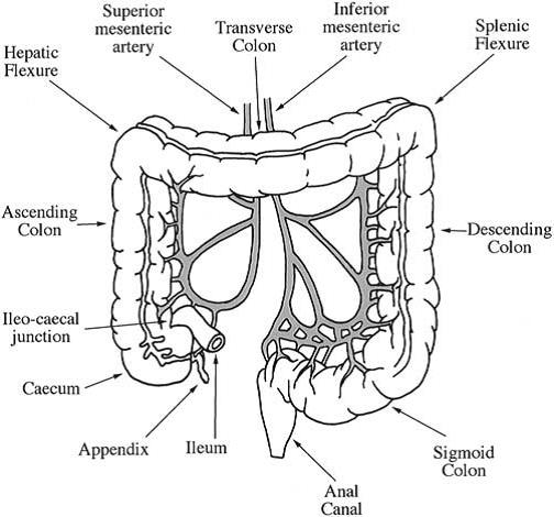
The GI tract is divided into stomach, small intestine and large intestine. The large intestine extending from the ileocaecal junction to the anus is divided into three main parts. These are the colon, the rectum and the anal canal. The location of the parts of the colon is either in the abdominal cavity or behind it in the retroperitoneum. The colon itself is made up of the caecum, the ascending colon, the hepatic flexure, the transverse colon, the splenic flexure, the descending colon and the sigmoid colon (Figure 1). It is about 1.5 m long, the transverse colon being the longest and most mobile part (Meschan, 1975), and has a average diameter of about 6.5 cm. The colon from the cecum to the splenic flexure (the junction between the transverse and descending colon) is also known as the right colon. The remainder is known as the left colon.

      Arterial supply to the colon of humans comes from branches of the superior and inferior mesenteric arteries. Venous drainage usually mirrors colonic arterial supply, with the inferior mesenteric vein draining into the splenic vein, and the superior mesenteric vein joining the splenic vein to form the portal vein, which then enters the liver.

       Lymphatic drainage from the entire colon and proximal two-thirds of the rectum is to the paraortic nodes, which then drain into the cisterna chyli. The lymph from the remaining rectum and anus can either follow the same route, or drain to the internal illiac and superficial inguinal nodes. The dentate line only roughly marks this transition.  

ANOTOMY & PHYSIOLOGY OF COLON

 Figure 1.  Main features of the colon

Colon – ANOTOMY AND PHYSIOLOGY OF COLON Functions Pharmacology Notes PDF DOC Colon – ANOTOMY AND PHYSIOLOGY OF COLON Functions Pharmacology Notes PPT Colon ANOTOMY PHYSIOLOGY OF COLON Functions Pharmacology Notes PDF ppt

Functions of Colon

The colon serves four major functions. They are

  1. Creation of suitable environment for the growth of colonic microorganisms
  2. Storage reservoir of faecal contents
  3. Expulsion of the contents of the colon at an appropriate time and
  4. Absorption of potassium and bicarbonate.


B Pharmacy M pharmacy Study Material Pharmacology Notes PDF DRUGS SUITABLE FOR COLONIC DRUG DELIVERY

B Pharmacy M pharmacy Study Material Pharmacology Notes DRUGS SUITABLE FOR COLONIC DRUG DELIVERY

B Pharmacy M pharmacy Study Material Pharmacology Notes is an article with detailed notes on DRUGS SUITABLE FOR COLONIC DRUG DELIVERY. 

 B Pharmacy M pharmacy Study Material Pharmacology Notes DRUGS SUITABLE FOR COLONIC DRUG DELIVERY

DRUGS SUITABLE FOR COLONIC DRUG DELIVERY

Drug delivery selectively to the colon through the oral route is becoming increasingly popular for the treatment of large intestinal diseases and for systemic absorption of protein and peptide drugs. There has been an increasing interest in utilizing the colon as a site for systemic absorption of these drugs in view of the less hostile environment prevailing in the colon. A variety of protein and peptide drugs like calcitonin, interferon, interleukins, erythropoietin and even insulin are being investigated for their absorption using colon specific drug delivery (Mackay and Tomlinson., 1993).

Inflammatory bowel disease (IBD) such as ulcerative colitis and Crohn’s disease require selective local delivery of drugs to the colon. Sulfasalazine is the most commonly prescribed drug for such diseases. Selective delivery of the drug to the colon is required for therapeutic efficacy with less or no side effects. The other drugs used in IBD are steroids, such as dexamethasone, prednisolone, and hydrocortisone.  In colonic cancer, anticancer drugs like 5-flurouracil, doxorubicin, and nimustine are to be delivered specifically to the colon. The site specific delivery of drugs like, metronidazole, mebendazole, albendazole is used in the treatment of infectious diseases, such as amoebiasis and helmenthiasis (Krishnaiah et al., 2002b; Krishnaiah et al., 2001; Jain et al., 2004).

DRUGS SUITABLE FOR COLONIC DRUG DELIVERY pharmacology study material notesClick here to download

{DOC} B Pharmacy M pharmacy Study Material Pharmacology Notes DRUGS SUITABLE FOR COLONIC DRUG DELIVERY PDF B Pharmacy M pharmacy Study Material Pharmacology Notes DRUGS SUITABLE FOR COLONIC DRUG DELIVERY

Besides peptide and protein drugs, the colon is also a good site for the absorption of drugs that are not stable in the acidic environment of the stomach, cause gastric irritation (e.g. aspirin, iron supplements) or those degraded by small intestinal enzymes. A number of drugs available as sustained release or delayed release or timed release tablets or capsules for oral administration are anti-inflammatory drugs, anti-hypertensive drugs, etc. Unless these drugs have good absorption characteristics in the colon, their intended use in the management of respective disorders through sustained release or timed release formulations will be in question.  The drugs that are having good absorption properties from the colon include theophylline, glibenclamide (Brockmeier et al., 1985), and oxprenolol (Devis et al., 1988). Diclofenac, ibuprofen, nitrendipine, isosorbide, metoprolol, nifedipine etc. and hence can be investigated for better bioavailability through colon specific drug delivery (Fara, 1989).


[Doc PDF PPT ] Adenosine: Pharmacology notes B pharm M pharmacy Study Material

Adenosine Pharmacology notes for B M Pharmacy students

Pharmacology noted for B pharmacy and M Pharmacy students is readily available in our site www.pharmawiki.in/ [Doc PDF PPT ] Adenosine: Pharmacology notes B pharm M pharmacy Study Material is here below. Have a look at it and study it.

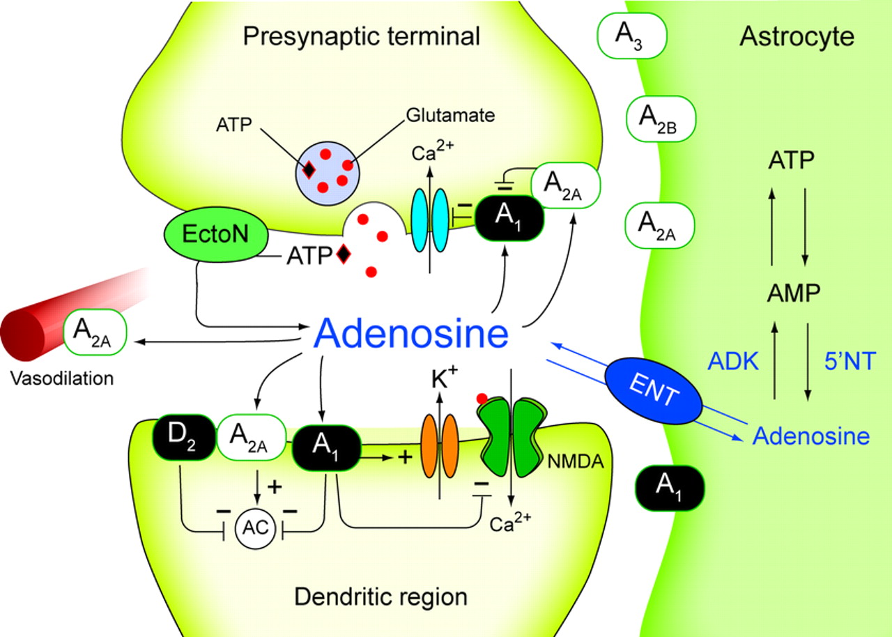
Adenosine is a purine nucleoside that regulates many physiological functions which includes respiratory regulation, neural function ,platelet aggregation, hormonal action , lymphocyte differentiation, vascular tone, negative chronotropic  and dromotropic effect on heart , also mediates inhibition of neurotransmitter release and lipolysis . These physiological function have been largely revised.(1),(2)

These functions are mediated through different adenosine receptor. There are four subtypes of AR-A1,A2A-AR,A2B-AR,A3-AR  each of these receptors has distinct tissue distribution and effector coupling. They belong to super family of G-protein coupled receptors (3).among these  receptors A1,A3AR1 are closely related  based on their sequence similarity while A2A,A2B AR also similarly related. A1 and A3 are primarly couple to G(subi) –family of G-protein.A2A and A2B are mostly coupled to GS  like G- protein. Each of these receptors plays an essential role in responding to adenosine in central nervous system(4) ,regulating pain (5) . cerebral blood flow(6). Basal gangalia function (7) respiration (8) and sleep (9.) thus these receptors can be therapeutic  targets for several diseases. Development of more selective agonists and antagonists  for adenosine receptor subtype provide aclass of therapeutics for treatment of numerous human diseases such as apain (10).  parkinsons disease (11)  asthma(12)   huntingtons disease(13).A search for new leads acting on specific adenosine acting on specific adenosine receptors may provide a key for novel therapeutics

Adenosine Pharmacology notes for B M Pharmacy students

Structure of A2A AR

        A2A-AR subtype is linked to  and G(S) and G(OLF) protein and up on activation the intracellular levels of Camp  are increased . the  expression  A2A AR expression is higest in brain, .spleen,thymus,leucocyte and blood platelets and intermediate in heart lungs and blood vessel.(14)(15)..Crystal structure of A2A AR was determined in 2008,physiological functions  A2A AR are regulation of sensori motors integration in basal ganglia., inhibition of platelet aggregation and polymorpho nuclear leucocytes, vasodilation protection  against ischemic damage, stimuation of sensory nerve activity . (17)  these wide range of functions implies their significant role in the body and use of chemical moieties to alter these function in disease state (may be agonists or antagonists).

A2A AR Adenosine antagonists:

A2A AR antagonist have their role in parkinsons disease, (18) keep regulations (19) controlling alcohol abuse (20) invivo receptor imaging (21)  there can also be used an anti depressant drug. (22) A2 AR agonists can be a treatment for ischemic renal injure (23) paraoxysmal supro ventricular tachycardia. They can be used as vasodilators (24) antithrombic agent (25)  antinflamatory (26) . they can also be used in treatment of asthma( 27), arthritis(28) sepsis (29) inflamatory bowel disease (30) and reduced skin pressure  ulcer formation (26) and accelerator  wound healing,(31)

In view of the role of A2A AR in these diseases afurther study in to the subject may reveal beneficial (facts) information for the treatment of such dieases. These receptors became agood targeting strategy  to bring out novel therapeutics for effective treatment of dieases.

Click here to download Pharmacology notes for B pharmacy M pharmacy StudentsADENOSINE

[DOC] Adenosine. Pharmacology notes B pharm M pharmacy study material

[Webpage] Adenosine. Pharmacology notes B pharm M pharmacy study material

[PDF] Adenosine. Pharmacology notes B pharm M pharmacy study material

Google Drive Link for Adenosine. Pharmacology notes B pharm M pharmacy study material

Pharmacology study material ADENOSINE Article REFRENCES:

(1) K. A. Jacoboson, Z.G.Gao, Adenosine receptors as therapeutic targets Nal.Rev., Drug Discovery 5(2006) 247-264

(2)M.P.Abbracchio, G.BUrnstock, A.verkhasatsky, H. Zimmermann, purinergic signalling in nervous system an over view ,Trends Neurosci. 32 (2009) 19-29

(3)B.B. Fredholm, G.Arsian, L.Halldner, B.kull, G.Schutte, W.Wasserman, structure and function of adenosine receptors and their genes , Naunyn – Schmiedebergs Arch.pnaarmacol.362 (2006) 19-29

(4)(a) . T.V Dunwiddie, S.A .Masina Annu . Rev.Neurosci 24,31(2001)

(b). K.A.Jacobson, Z.G.Gao , Nat .Rev Drug Discover.5,247 (2006)

(5) J.sawynok,x.J.Liv,Drog Neurobio . 69,313 (2003)
(6) Y.Shietal, J.Cereb Blood Flow Method . 28,111 (2008)

(7) M.A.Schwarzchild , L.Agnati, K. Fure, J. Fichsn, M.M orelli , Trends Neurobiol 29. 647 (2006)

(8) S.Lahiri, CH.Nitchell.D.Reigade , A.Roy , N.s.chemiack , Respir. Physiol. Nenrobiol. 157 ,123 (2007)

(9) R.Basheer,R.E.Strecker,MM.Thatkar,R.W.M.Carley Prog. Neurobiol.73,379 (2009)

(10)J,Sawynok,X.J.Liu. Prpg.Neurobio.69,313

(11)A.H.Schapira.et.al, Nat.Rev.Drug.Discor.5,845 (2006)
(12)A. Brow, D.Spina, C.p.page, Br.J.Pharmacol.153,(suppli),5446(2008)

(13)D.Blum, R.Hourez, M.C.Galar, P.Popoli,S.N.Schiftmann, Lancet Neurol.2,366(2003)

(14)F.Meng, G.X.Xic, D.Chalmeri, C.Margan,S.J.Watson,Jr.,H.Akil,Cloning and expression of the A2a  receptors from guinea pig brain Neuro chem 66(1996) 613-621

(15)R.A.Deter freud,M.Maccollin,J.Gusella,J.S.Flink Characterization and expression of the human A2a adenosine receptors gene, Neuro chem. 66(1996)362-368.

(16)Veli-Pekka Jaakola, Mark.T.Grifftin,Micheal, A.Hanson, Vadim cherezov , ellen y.t chien, J.Robert lane ,Adlioan, P.L.Jzerman, Raymond c.sterenes, the 2.6 Angstroun Crystal structure of a human A2a Adenosine  receptor Bound to an Antagonist

(17)B.B.Fredholm, Adenosine, an endogenous distress signal, modulates tissue damage and repair not cell death and differentiation (2007) 14, 1315-1323

(18)Michael A .Schwarzschild, Luigi Agnati, Kjell Fuxe ,Jiang – fanchen and micaela morelli, Targetting Adenosine A2a receptors in parkinsons disease Trends in neurosciences Vol.29 No.11

(19) Satoh, S., Matsumura, H. & Hayaishi, O. Involvement of adenosine A2A receptor in sleep promotion. Eur. J. Pharmacol. 351, 155–162 (1998

(20) Yao, L. et al. dimers mediate synergy of dopamine D2 and adenosine A2 receptor-stimulated PKA signalling and regulate ethanol consumption. Cell 109, 733–743 (2002).

(21) Moresco, R. M. et al. In vivo imaging of adenosine A2A receptors in rat and primate brain using [11C]SCH442416. Eur. J. Nucl. Med. Mol. Imaging 32,405–413 (2005).

(22) El Yacoubi, M. et al. Absence of the adenosine A2A receptor or its chronic blockade decrease ethanol withdrawal-induced seizures in mice. Neuropharmacology 40, 424–432 (2001).

(23) Okusa, M. D. et al. A2A adenosine receptor-mediated inhibition of renal injury and neutrophil adhesion. Am.J. Physiol. Renal Physiol. 279, F809–F818 (2000).

(24) Fredholm, B. B., IJzerman, A. P., Jacobson, K. A., Klotz, K. N. & Linden, J. International Union of Pharmacology. XXV. Nomenclature and classification of adenosine receptors. Pharmacol. Rev. 53,527–552 (2001).

(25) Varani, K. et al. Dose and time effects of caffeine intake on human platelet adenosine A2A receptors:functional and biochemical aspects. Circulation 102,285–289 (2000)

(26) Peirce, S. M., Skalak, T. C., Rieger, J. M.,Macdonald, T. L. & Linden, J. Selective A2A adenosine receptor activation reduces skin pressure ulcer formation and inflammation. Am. J. Physiol. Heart Circ. Physiol. 281, H67–H74 (2001).

(27) Fozard, J. R., Ellis, K. M., Villela Dantas, M. F., Tigani, B. & Mazzoni, L. Effects of CGS 21680, a selective adenosine A2A receptor agonist, on allergic airways inflammation in the rat. Eur. J. Pharmacol438, 183–188 (2002).

(28) Montesinos, M. C. et al. Adenosine A2A or A3 receptors are required for inhibition of inflammation by methotrexate and its analog MX-68. Arthritis Rheum. 48, 240–247 (2003

(29) Sullivan, G. W., Fang, G., Linden, J. & Scheld, W. M. A2A adenosine receptor activation improves survival in mouse models of endotoxemia and sepsis. J. Infect. Dis. 189, 1897–1904 (2004).

(30) Odashima, M. et al. Activation of A2A adenosine receptor attenuates intestinal inflammation in animal models of inflammatory bowel disease. Gastroenterology 129, 26–33 (2005).

(31) Montesinos, M. C. et al. Wound healing is accelerated by agonists of adenosine A2 (Gs-linked) receptors. J. Exp. Med. 186, 1615–1620 (1997).